Catégories
Armes electroniques General

Une expérience de stimulation (TMS) cérébrale soulage la dépression


Tu ne supportes plus qu’on tue/asservisse/torture… l’humain en masse partout dans le monde, tu ne supportes plus la marche forcée que t’impose le monde.
Tu es en dépression mais on va te rendre compatible avec ce cauchemar ! Tu seras heureux, tu pourras enfin t’intégrer dans cette société malade. Une vraie thérapeutique Orwellienne digne du meilleur soma. Et en plus, on peut même te rendre heureux à distance et potentiellement à « l’insu de ton plein gré » (une véritable fabrique de psychopathes), comme le démontre le brave Dr James Giordano à l’académie militaire de West Point. Pendant ce temps, au lieu de s’unir, les noirs s’opposent aux « privilèges blancs » dans un combat d’un autre siècle (je ne dis pas inutile), suite au décès de George Floyd (et ici en France avec adama Traoré) qui n’a pas été traité avec les égards qu’aurait dû lui assurer la démocratie (il y a des vidéo de blancs qui meurent de la même manière sur internet). Ce qu’il faut bien comprendre avec de telles armes, toujours présentées par les médias comme avancée médicale positive, c’est que peu importe la couleur de peau, nous sommes 99% à avoir le profil pour devenir les « nègres » du 21 siècle. Il y a plus urgent et actuel que les guerres raciales horizontales, il est temps de comprendre(et d’agir en sorte) que l’élite dirigeante et financière mène la guerre au peuple quelle que soit sa couleur de peau.


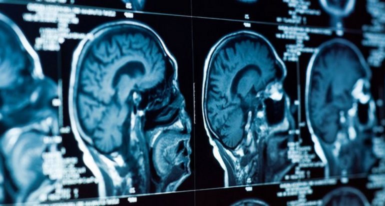
Stimuler des zones clés du cerveau avec un champ magnétique pulsé pourrait représenter une solution pour certaines personnes souffrant de dépression chronique qui n’a pas pu être traitée à l’aide d’autres techniques ou médicaments conventionnels.

Les résultats très encourageants d’une expérience effectuée sur un petit groupe de volontaires suggèrent qu’un traitement stimulant certaines parties clés du cerveau avec un champ magnétique pulsé pourrait soigner la dépression chronique chez certaines personnes qui souffrent d’une dépression résistante à d’autres traitements conventionnels.

La base de ce traitement non invasif, appelé stimulation intermittente par rupture thêta (iTBS), a déjà été approuvée comme thérapie pour la dépression grave par la U.S. Food and Drug Administration (FDA).

Des chercheurs de l’Université de Stanford et de l’Université de Palo Alto aux États-Unis ont montré, lors d’un essai effectué sur 21 personnes, qu’administrer cinq fois la dose globale de pulsations sur un nombre plus élevé de séances quotidiennes semble non seulement sûr, mais pourrait permettre d’obtenir de bien meilleurs résultats.

Des impulsions magnétiques pour établir de meilleures connexions entre les neurones
Ce traitement implique de diriger des centaines d’impulsions magnétiques intenses dans une partie spécifique du cerveau, durant quelques minutes par jour pendant six semaines, ce qui permet d’encourager les neurones à établir lentement des connexions plus solides qui peuvent aider à atténuer les symptômes de la dépression chronique. Cette procédure s’est avérée suffisamment efficace pour donner de l’espoir aux 10 à 30% des patients souffrant de dépression, qui ne répondent pas facilement aux autres thérapies.

Une étude publiée en 2018 a révélé qu’un peu moins de la moitié des personnes diagnostiquées avec une dépression ont montré au moins une certaine amélioration après ce traitement, avec environ un tiers des individus en rémission. Et lorsque rien d’autre ne fonctionne, il s’agit de chiffres plutôt attrayants.

Cependant, les chances de succès pourraient être meilleures, d’autant plus que la tentative de stimulation magnétique transcrânienne n’est pas nécessairement exempte d’effets secondaires, qui peuvent inclure des maux de tête et d’autres inconforts légers. Cette dernière étude suggère fortement que de bons résultats sont possibles, mais il est important de garder à l’esprit que l’expérience a été menée sur une très petite quantité d’individus, et sur une très courte durée. D’autant plus que l’étude n’avait pas de groupe témoin pour mesurer les résultats.

Après cinq jours comprenant 10 séances iTBS à haute dose par jour, 19 des 21 volontaires ont exprimé une amélioration suffisante de leur dépression, pour compter comme étant en rémission.

Les chercheurs estiment que si les essais continuent à démontrer de tels résultats positifs, cette technique pourrait bien un jour aider jusqu’à 90% des personnes souffrant de dépression. « Il n’y a jamais eu de théra­pie pour les dépres­sions résis­tantes aux trai­te­ments qui ont présenté plus de 55% de rémis­sion dans des condi­tions simi­laires », a expliqué le psychiatre de Stanford, Nolan Williams.

Le trouble dépressif majeur est une maladie complexe que nous n’avons pas encore pleinement comprise et qui peut être provoquée par de nombreux facteurs (biologique, psychologique, social, fonctionnement du cerveau), qui ne sont eux-mêmes pas encore bien compris. Selon les chercheurs, une fraction de ces derniers ne pourrait pas être traitée à l’aide de produits pharmaceutiques conventionnels.

Source

Catégories
Armes electroniques

Neurosciences – Armes à Energies Dirigées, Dr Giordano, à l’académie militaire de West Point.

MK ULTRA – Dr James Giordano – Neurosciences – Armes à Energies Dirigées – Nano Implants – 2018

Traduction faite par Nicocible, d’une conférence du Dr Giordano, à l’académie militaire de West Point.

Nicociblé :

Son Site

Sa chaine Youtube


Dans cette vidéo, le Dr James Giordano parle aux cadets et aux professeurs de l’Académie militaire américaine du cerveau humain et de l’avenir de la guerre. Giordano est chef du programme d’études en neuroéthique et chercheur au Centre Pellegrino de bioéthique clinique de l’université de Georgetown. L’histoire récente a vu des avancées substantielles dans les domaines des neurosciences et de la neurotechnologie, explique Giordano. Il est presque certain que ces progrès auront un impact sur les guerres qui seront menées à l’avenir. À bien des égards, le cerveau pourrait même faire partie de l’espace de combat. Cet événement a été organisé par l’Institut de la guerre moderne.


Un passage important en capture d’écran

Catégories
Armes electroniques

Puis ils sont venus pour votre esprit : L’histoire inédite de la psychochirurgie

SOURCE

Peter et Ginger Breggin – est l’interview complète, brute et encore inédite, du Dr Breggin qui a été utilisée dans notre film « The Minds of Men ».

Le Dr Breggin rend compte de son activisme et de sa lutte contre la psychochirurgie il y a plus de 50 ans – une campagne des années 1960-70 visant à légitimer la destruction du cerveau et à utiliser un mode d’intervention dans le cerveau en constante évolution et de plus en plus technologique, utilisant des électrodes implantées pour cibler des zones minuscules du cerveau liées au comportement, et pour utiliser la surveillance et l’activation par radiofréquence (RF) pour contrôler les humeurs, l’attitude et peut-être même la pensée.

Pire encore, les praticiens et les décideurs qui ont poussé cette nouvelle ère dangereuse d’intervention dans notre espace mental privé le faisaient sur la base de la désactivation PRÉEMPTIVE de ceux qui étaient identifiés comme « potentiellement violents ». Comme nous l’avons expliqué dans « L’esprit des hommes », une campagne massive était en cours à la suite de fusillades et d’assassinats de masse pour filtrer le public et attirer un large éventail de personnes qu’il souhaitait pacifier et sortir de l’équation.

Il s’agissait de militants et de dirigeants politiques de la communauté noire et d’autres fauteurs de troubles potentiels – des responsables politiques, et non des délinquants ou des personnes qui avaient « fait quelque chose de mal ». Le programme a également ciblé les personnes « à risque » dans toutes les couches de la société.

Le Dr Breggin a joué un rôle essentiel en braquant les projecteurs sur ces pratiques gênantes – en mettant l’accent sur les travaux du Dr Jose Delgado et des docteurs Vernon Mark, Frank Ervin et William Sweet (également représentés dans « The Minds of Men »).

Se dressant contre cette injustice, Breggin a résisté aux menaces de mort et à l’opposition des milieux psychiatriques et psychologiques dans lesquels il a été formé pour mettre fin au financement et à la pratique – du moins ouvertement.

Ce qui a continué dans l’ombre est une autre histoire… mais une histoire qui se rattache solidement à l’endroit que la société a de nouveau atteint – la croyance dangereusement naïve que les « autorités », les « experts », les « milliardaires » et leurs technologies peuvent non seulement améliorer le monde, mais aussi réparer nos cerveaux et nos esprits.

C’est une histoire importante qui était, jusqu’à présent, peu connue.

Le Dr Breggin et la lutte contre la psychochirurgie sont présentés dans « The Minds of Men » :


Complimenting yesterday’s post – a guest article written by Dr. Peter & Ginger Breggin – is the raw, and as yet unreleased full interview with Dr. Breggin that was used in our film “The Minds of Men”.

Dr. Breggin gives an account of his activism and struggle against psychosurgery more than 50 years ago – a 1960s-70s era campaign to legitimize the destruction of the brain, and utilize an evolving and increasingly technological mode of intervention in the brain using implanted electrodes to target minute areas in the brain tied to behavior, and to use radio frequency (RF) monitoring and activation to control moods, attitude and perhaps even thought.

Worse, the practitioners and agenda-makers who were pushing this dangerous new era of intervention in our private mind space were doing so on the basis of PREEMPTIVELY disabling those identified as ‘potentially violent.’ As we detailed in ‘The Minds of Men,’ a massive campaign was underway in the wake of mass shootings and assassinations to screen the public, and draw a wide net of people they wanted to pacify and take out of the equation.

These included activists and political leaders in the black community and other potential troublemakers – political liabilities, not offenders or those who’d ‘done something wrong.’ The program also targeted “at risk” individuals throughout the strata of society.

Dr. Breggin played a pivotal role in shining a spotlight on these troublesome practices – with a focus on the work of Dr. Jose Delgado, and doctors Vernon Mark, Frank Ervin and William Sweet (also depicted in “The Minds of Men”).

Standing up against this injustice, Breggin withstood threats on his life and opposition from within the psychiatric and psychological fields which he was trained in to stop the funding and practice – at least overtly.

What continued in the shadows is another story… but one which connects solidly with the place society has once again reached – the dangerously naive belief that ‘authorities’ and ‘experts’ and ‘billionaires’ and their technologies can not only improve the world, but fix our brains and minds as well.

This is an important story that has been, until now, little known. Please watch and share.

Dr. Breggin and the fight against psychosurgery is featured in “The Minds of Men”:

Catégories
Armes electroniques

Armes bio-électromagnétiques : L’arme ultime

Armes bio-électromagnétiques : L’arme ultime

Bio-electromagnetic Weapons: The ultimate weapon  

Un système d’armes qui fonctionne à la vitesse de la lumière, qui peut tuer, torturer, asservir et échapper à la détection, Harlan Girard

L’arme ultime

Les armes électromagnétiques fonctionnent à la vitesse de la lumière ; elles peuvent tuer, torturer et asservir ; mais le public ignore largement leur existence, car ces armes fonctionnent de manière furtive et ne laissent aucune preuve physique. Les armes électromagnétiques ont été testées sur des êtres humains depuis 1976. En dispersant largement les sujets de ces tests humains involontaires et en attaquant avec véhémence leur crédibilité, les États-Unis ont pu procéder à ces expériences humaines sans être gênés par des discussions ou des critiques, et encore moins par une opposition.

Ce système d’arme ultime est actuellement déployé en Irak. L’US Air Force et le Marine Corps l’appellent « technologie de déni actif », comme si elle était utilisée uniquement à des fins de défense, mais ce n’est pas le cas (voir encadré 1).

La vérité sur la « technologie du déni actif ».

Il n’y a qu’un seul spectre électromagnétique. Les armes nucléaires libèrent une grande quantité de rayonnements ionisants dans la gamme des hautes fréquences au-dessus de la lumière visible, où l’énergie des rayonnements est capable de rompre les liaisons chimiques. Il est généralement reconnu que les rayonnements ionisants peuvent provoquer le cancer.

L’armée américaine a transformé en armes les radiations non ionisantes en dessous de la gamme visible, les micro-ondes et les ondes radio qui sont utilisées dans les téléphones mobiles et les télécommunications. Le gouvernement américain a vigoureusement nié que les radiations électromagnétiques non ionisantes puissent présenter des risques pour la santé, à la fois pour défendre la recherche sans consentement sur l’homme qu’il mène depuis de nombreuses années mais qu’il n’a pas encore reconnue, et pour dissuader d’autres pays de développer des armes similaires.

Le seul effet biologique des radiations non ionisantes que le gouvernement américain a reconnu depuis de nombreuses années est le réchauffement et, par conséquent, il caractérise la « technologie de déni actif » comme celle qui produit des douleurs dues à un réchauffement soudain de la peau ; mais ce n’est pas ainsi que cela fonctionne réellement.

Lecture des ondes cérébrales et contrôle de l’esprit

En 1959, Saul B. Sells, professeur de psychologie sociale dans une petite université américaine, a soumis une proposition à la Central Intelligence Agency (CIA) pour construire pour eux l’appareil d’électroencéphalographie le plus sophistiqué qui aurait une capacité de calcul intégrale pour analyser et, espérons-le, donner un sens aux ondes cérébrales qu’il enregistre. En d’autres termes, le professeur a proposé de fabriquer une machine qui pourrait dire à la CIA ce qu’une personne pense, qu’elle souhaite ou non divulguer cette information.

La CIA a approuvé le projet en 1960, en y ajoutant des recherches en bibliothèque avec cinq objectifs. Le cinquième objectif de la recherche était, « Techniques d’activation de l’organisme humain par des moyens électroniques à distance ». L’ensemble de cette mission a ensuite été connu sous le nom de sous-projet MKULTRA 119, MKULTRA étant le célèbre programme de contrôle des esprits de la CIA. Il était basé sur l’idée erronée que les Soviétiques possédaient déjà les moyens de contrôler les esprits et que les États-Unis devaient rattraper leur retard le plus rapidement possible.

Les documents relatifs au sous-projet 119 de MKULTRA sont maintenant conservés aux National Security Archives (une organisation non gouvernementale) de l’Université George Washington à Washington, D.C. [1]. John Marks, auteur de The Search for the Manchurian Candidate : The CIA and Mind Control (Times Books, New York, 1979) a fait don des documents MKULTRA ; son livre a été réédité par W.W. Norton & Company, Inc, New York, 1991 et est toujours en cours d’impression.

Le projet Bizarre a suivi le sous-projet 119 de MKULTRA en 1965. Le but de Bizarre était d’enregistrer et d’analyser le signal micro-ondes complexe qui aurait été émis à l’ambassade américaine à Moscou par les Soviétiques depuis un bâtiment situé de l’autre côté de la rue. Ce qui est intéressant dans le projet Bizarre, c’est que si les États-Unis ont nié jusqu’à ce jour qu’il puisse y avoir des effets néfastes sur la santé à cause du rayonnement micro-ondes, ils ont immédiatement suspecté que « le signal de Moscou » produisait divers effets sur la santé du personnel de l’ambassade, en particulier chez les ambassadeurs successifs au bureau desquels il était prétendu que le signal était émis. Au moment même où le Département d’État testait le personnel de l’ambassade pour détecter les cassures d’ADN produites par le signal de Moscou, il s’est senti contraint de ne pas se plaindre aux Soviétiques parce que la puissance de leur signal était une infime fraction de ce que les États-Unis considéraient comme un niveau d’exposition humaine sans danger. Le journaliste Barton Reppert a écrit le compte-rendu le plus fiable sur le signal de Moscou [2]. (Note de la rédaction : les ruptures d’ADN dues à l’exposition aux téléphones portables ont été confirmées par des recherches récentes en laboratoire [3, 4] (Science in Society 24)).

Conversion du son aux micro-ondes

En 1973, Joseph C. Sharp, un psychologue expérimental de l’Institut de recherche de l’armée Walter Reed, a réalisé une expérience qui a été déterminante pour le développement de l’équipement de torture qui est aujourd’hui expédié en Irak. Il a fait installer dans son laboratoire par James Lin un équipement qui convertissait la forme des ondes sonores en un rayonnement micro-ondes qui lui permettait de s’entendre vocaliser les noms des nombres de un à dix dans sa tête, en contournant le mécanisme de ses propres oreilles. Cette expérience particulière n’a jamais été publiée mais est mentionnée dans le livre de Lin, Microwave Auditory Effects and Applications, publié en 1978 [5].

L’expérience a été confirmée par le brevet américain 6 587 729, « Apparatus for Audibly Communicating Speech Using the Radio Frequency Hearing Effect » [6] . Ce brevet concerne une version améliorée de l’appareil utilisé lors de l’expérience en laboratoire de 1973, délivrée le 1er juillet 2003 et attribuée au secrétaire de l’armée de l’air. Il apporte la preuve scientifique qu’il est possible d’entendre des voix menaçantes dans sa tête sans souffrir de schizophrénie paranoïaque.

Pourquoi ce brevet a-t-il été publié ouvertement à un moment où le gouvernement américain pratique un degré de secret qui rivalise avec le Kremlin de Staline ? Je n’ai pas de réponse satisfaisante, si ce n’est que l’appareil faisant l’objet du brevet a déjà été remplacé par un équipement qui permet d’obtenir le même effet par des moyens beaucoup plus sophistiqués. Il bloque les processus normaux de mémoire et de pensée par des moyens électroniques à distance, tout en fournissant des souvenirs et des suggestions fausses, déformées et/ou désagréables par un processus appelé « télépathie synthétique ». L’équipement qui produit la télépathie synthétique est parfois appelé « technologie d’influence ».

Si les voix et les visions, les rêves et les cauchemars sont les manifestations les plus étonnantes de ce système d’armes, il est également capable de paralyser le sujet humain en limitant son amplitude de mouvement normale, en provoquant une douleur aiguë équivalente à la défaillance d’un organe majeur, voire à la mort, et en interférant avec le fonctionnement normal de l’un des sens humains. En d’autres termes, toutes les tortures dont les mots Guantanamo Bay sont devenus synonymes peuvent être réalisées par des moyens électroniques à distance.

Instruments de torture

La technologie d’influence est également capable de persuader les sujets que leur esprit est lu, que leur propriété intellectuelle est pillée, et peut même motiver le suicide ou le meurtre de leur famille, de leurs amis et de leurs collègues. Pendant les années de la « guerre contre la drogue » (qui a précédé la « guerre contre le terrorisme »), les lettres que les sujets humains « involontaires » avaient écrites ou étaient sur le point de recevoir disparaissaient régulièrement du courrier, comme si le gouvernement menait une vaste opération secrète par l’intermédiaire des bureaux de poste du pays. Lorsque George Herbert Walker Bush est devenu président (en 1989), l’incidence des meurtres de collègues dans les bureaux de poste est devenue si importante que l’expression « going postal » a commencé à remplacer l’expression couramment utilisée de « going crazy ». Les meurtres de collègues sur d’autres lieux de travail ont également commencé à attirer l’attention des médias [7].

J’estime que le coût de l’emprisonnement d’un être humain dans son propre corps et de l’application d’une torture incessante est de 5 000 000 à 10 000 000 $US par an (voir ci-dessous).

C’est exactement ce que j’entends par « torture permanente ». Comme ce nouveau matériel de torture ne laisse aucune trace visible, telle que des lésions cutanées, il est possible de torturer les sujets humains involontaires 24 heures sur 24, 365 jours par an. Cela peut être fait et est fait même à Noël et à Pâques [8].

Je suis arrivé à mon estimation du coût des tests/utilisation d’armes électromagnétiques sur un sujet humain en visitant une chaîne de télévision câblée spécialisée dans la vente de marchandises par voie hertzienne 24 heures sur 24. Je me suis interrogé sur le nombre de techniciens nécessaires, leurs horaires de travail et leur fourchette de salaire, ainsi que sur le nombre de personnel d’appoint nécessaire pour préparer la programmation en vue de sa diffusion. Je ne me suis pas enquis du coût de l’électronique et de la grille d’amortissement. J’ai estimé un coût de dépréciation qui est inclus dans mon estimation du coût de la torture d’un sujet humain « involontaire » pendant un an.

La torture est une activité à forte intensité de main-d’œuvre. Quel objectif justifierait cet investissement ? Serait-ce quelque chose d’aussi insensé que de gouverner le monde en asservissant les gouvernements démocratiques des pays les plus peuplés ? Cet objectif est certainement cohérent avec le mépris et l’hostilité des États-Unis envers les Nations unies, les conventions et les pactes internationaux qu’ils ont ratifiés par le passé et le droit international coutumier [9].

Le 1er mars 2001, le Corps des Marines a annoncé une nouvelle arme non létale, la « technologie de déni actif ». Elle produit une douleur énorme en faisant soi-disant bouillir les molécules d’eau de la peau humaine sans endommager la peau elle-même. Comme décrit dans un article publié dans le New Scientist, elle utilise un rayonnement électromagnétique pulsé à une fréquence de 95 GHz avec une portée d’environ 600 mètres [10]. Plusieurs nouveaux rapports ont été publiés dans le magazine en 2005, dont un en juillet [11], décrivant des volontaires participant à des tests visant à déterminer le degré de sécurité de l’arme Active Denial System (ADS) si elle était utilisée dans le cadre d’un véritable contrôle de foule. Il a été rapporté que le faisceau de l’arme ADS provoque une douleur en 2 à 3 secondes, et devient « intolérable après moins de 5 secondes ».

La technologie de déni actif est la pierre angulaire du système utilisé pour torturer 2 000 personnes dans l’intimité de leur foyer, non seulement aux États-Unis mais dans le monde entier, partout où des pays ont signé des accords sur le statut des forces avec les États-Unis [12]. Les premières allégations de torture ont été reçues de pays avec lesquels les États-Unis ont une relation spéciale de partage de renseignements, à savoir le Royaume-Uni, le Canada, l’Australie et la Nouvelle-Zélande. Puis des rapports ont commencé à arriver des pays conquis où les États-Unis ont encore un grand nombre de troupes stationnées, c’est-à-dire l’Allemagne et le Japon. Lorsque la France a rejoint le bras militaire de l’OTAN à la fin des années 90, nous avons commencé à recevoir des allégations de torture en France. Très récemment, nous avons commencé à recevoir des allégations de torture en Inde, où des entreprises américaines ont commencé à externaliser, non seulement les lignes d’assistance, mais aussi la programmation [13].

Et puis il y a le cas de la Russie, où les sujets humains « involontaires » d’expériences de torture semblent être à la fois nombreux et bien organisés. On m’a dit avec certitude que tous les scientifiques russes qui parlaient anglais ont maintenant trouvé un foyer dans une université américaine ou un laboratoire gouvernemental. C’est plausible, si l’on considère la crainte fréquemment exprimée par les Américains que les experts de l’ère soviétique en matière d’armes nucléaires et de guerre biologique ne trouvent un emploi en Iran. Il est un fait qu’à la fin de la Seconde Guerre mondiale, l’armée américaine a balayé l’Allemagne dans une opération appelée « Project Paperclip », recrutant notamment des spécialistes des fusées nazies et des experts en médecine aérospatiale. D’autres scientifiques ont été recrutés simplement pour priver l’Union soviétique de cette ressource. Qu’est-il donc advenu des scientifiques soviétiques qui ne parlaient pas anglais ? Nous le saurons avec le temps, mais pour l’instant, on peut supposer qu’au moins certains d’entre eux ont été employés pour étudier le système de valeurs et les processus de décision russes en torturant d’autres Russes avec la « technologie d’influence » américaine [14].

Deux articles intéressants et importants sur les armes bioélectromagnétiques ont récemment été publiés dans le New Scientist : « La nouvelle arme américaine vise la douleur maximale » et « Police toy with ‘less lethal’ weapons », tous deux écrits par David Hambling [15]. Voir aussi US. Brevet 6 536 440 du 25 mars 2003 [16].

Depuis la fin de cet article à la mi-juin 2005, il a été porté à mon attention que les Israéliens déploient un dispositif appelé « The Scream », qui émet des rafales de sons audibles, mais pas forts, à des intervalles d’environ 10 secondes. Un photographe présent sur les lieux d’une manifestation a déclaré qu’il continuait à entendre le son dans sa tête même après s’être couvert les oreilles. Cela me fait penser que l’agent actif est électromagnétique plutôt qu’acoustique. En d’autres termes, les Israéliens ont mis au point un dispositif qui est bien plus intelligent que notre « technologie de déni actif ». Non seulement il dissuade les émeutiers, mais il émet également un avertissement sonore indiquant qu’il a été activé, ce qui n’est pas le cas du dispositif américain, laissant aux hommes enrôlés qui l’utilisent le soin de déterminer l’intensité des brûlures que reçoivent leurs adversaires. La marge d’erreur du dispositif américain est déraisonnable. On peut tout aussi bien l’appeler une arme mortelle car, dans la pratique, elle le sera très fréquemment [17].

La source originale de cet article est le communiqué de presse de l’ISIS

Copyright © Institut des sciences dans la société, Communiqué de presse ISIS, 2007

Catégories
Armes electroniques

La mise au point de nouvelles armes antipersonnel – Revue internationale de la Croix-Rouge

N°2 de la traduction d’articles issus du site MindJustice. « Sample of Rare and Outstanding Articles from 1976 to 1996 »

International Review of the Red Cross, 279

November 1, 1990
The Development of New Antipersonnel Weapons
by Louise Doswald-Beck and Gerald C. Cauderay

***

Les blessures qui en résultent sont aujourd’hui, et le seront dans un avenir prévisible, pratiquement inexistantes. Le deuxième groupe de travail, composé principalement de psychiatres et de médecins, étudiera plus en détail les effets à court et à long terme, tant pour l’individu que pour la société, de la cécité &a par rapport aux autres blessures généralement subies sur le champ de bataille. Les informations recueillies peuvent ensuite être utilisées pour une discussion plus approfondie sur les implications juridiques et politiques de la mise au point de ces armes.

5. Armes à énergie dirigée (DEW)

Outre l’arme laser antipersonnel, qui à certains égards pourrait également être considérée comme une arme à énergie dirigée, il existe également des armes très spéciales, telles que celles utilisant des ondes électromagnétiques de différentes longueurs d’onde et des générateurs de faisceaux de particules, qui sont considérées par certains experts comme des armes antimatérielles potentielles extrêmement efficaces. S’il est peu probable que ce type particulier d’arme, qui nécessite un approvisionnement énergétique considérable, devienne opérationnel sur le champ de bataille dans un avenir proche, il n’en va pas de même pour les systèmes d’armes utilisant des faisceaux d’ondes ou d’impulsions électromagnétiques. Les effets induits chez l’homme par les ondes électromagnétiques sont connus, quoique imparfaitement, depuis longtemps et ont fait l’objet de recherches continues. Selon la fréquence utilisée, le mode d’émission, l’énergie rayonnée, la forme et la durée des impulsions utilisées, les rayonnements électromagnétiques dirigés contre le corps humain peuvent produire de la chaleur et provoquer de sérieux problèmes ou même des changements dans la structure moléculaire des cibles qu’ils atteignent.

Des travaux de recherche dans ce domaine ont été menés dans presque tous les pays industrialisés, et notamment par les grandes puissances, en vue d’utiliser ces phénomènes à des fins antimatérielles ou antipersonnel. Des tests ont démontré que de puissantes impulsions micro-ondes pouvaient être utilisées comme une arme pour mettre l’adversaire hors de combat ou même le tuer. Il est possible aujourd’hui de générer une impulsion hyperfréquence très puissante (par exemple entre 150 et 3 000 mégahertz), avec un niveau d’énergie de plusieurs centaines de mégawatts. Grâce à des systèmes d’antennes spécialement adaptés, ces générateurs pourraient en principe transmettre sur des centaines de mètres suffisamment d’énergie pour cuisiner un repas. Toutefois, il est important de mentionner que les effets mortels ou incapacitants auxquels on peut s’attendre des systèmes d’armes utilisant cette technologie peuvent être produits avec des niveaux d’énergie beaucoup plus faibles. Utilisation du principe de concentration du champ magnétique, qui permet le contrôle de la géométrie de la cible, au moyen de systèmes d’antennes spécialement conçus à cet effet,

l’énergie rayonnée peut être concentrée sur de très petites surfaces du corps humain, par exemple la base du cerveau où une énergie relativement faible peut produire des effets mortels. Il semble qu’avec la technologie actuellement disponible, on pourrait sérieusement envisager la production de tels systèmes d’armes, qui pourraient avoir une portée d’environ 15 km et balayer une zone avec une série d’impulsions rapides. Les soldats non protégés dans cette zone pourraient être mis hors de combat ou tués en quelques secondes. Une telle arme pourrait être installée sur un camion et serait donc facilement transportable.

Malgré la rareté des publications sur ce sujet, et le fait qu’il s’agit généralement d’informations strictement classifiées, les recherches menées dans ce domaine semblent avoir démontré que de très faibles quantités de rayonnement électromagnétique pouvaient altérer sensiblement les fonctions des cellules vivantes. Les travaux de recherche ont également révélé que des effets pathologiques proches de ceux induits par des substances hautement toxiques pouvaient être produits par des rayonnements électromagnétiques même à très faible puissance, en particulier ceux utilisant une forme d’impulsion contenant un grand nombre de fréquences différentes. Comme mentionné précédemment, l’énergie nécessaire pour atteindre ces résultats est souvent beaucoup plus faible que l’énergie nécessaire pour induire un effet significatif de la chaleur dans les tissus du corps.

Certaines recherches semblent avoir confirmé que les champs électromagnétiques de faible intensité, modulés de façon à être similaires aux ondes cérébrales normales, pourraient sérieusement affecter les fonctions cérébrales. Des expériences avec des champs magnétiques pulsés effectuées sur des animaux auraient produit des effets spécifiques tels que l’endormissement et le déclenchement de l’anxiété ou de l’agressivité, en fonction de la modulation de la fréquence utilisée. D’autre part, il est bien connu que des effets mortels peuvent également être produits en utilisant des niveaux de puissance plus élevés que ceux utilisés pour les expériences sur la modification du comportement. Une arme antipersonnel basée sur de tels principes biophysiques pourrait produire des effets similaires à ceux d’un gaz neurotoxique, mais n’aurait aucun effet secondaire et ne laisserait aucune trace durable.

***

International Review of the Red Cross, 279

November 1, 1990
The Development of New Antipersonnel Weapons
by Louise Doswald-Beck and Gerald C. Cauderay

17 of 27
resulting injuries is at present, and will be for the foreseeable future, virtually non-existent. The second working group, which will principally comprise psychiatrists and doctors, will study in greater detail the short and long-term effects, both  for the individual and for society, of blindness &a compared with other injuries typically sustained on the battlefield.  The information collected can then be used for a more thorough discussion of the legal and policy implications of the development of these weapons.
5. Directed energy weapons (DEW)
Apart from the anti-personnel laser weapon, which in some respects could also be considered as a directed energy weapon, there are also very special weapons, such as those using electromagnetic waves of different wavelengths   and generators of particle beams, which are considered by some experts as extremely efficient potential anti-materiel weapons. Although this particular type of weapon, which requires a considerable energy supply, in unlikely to become operational on the battlefield in the near future, the same cannot be said for weapon systems using beams of electromagnetic waves
18 of 27
or pulses. The effects induced in human beings by electromagnetic waves have been known, albeit imperfectly, for a long time and have been the subject of continuous research. Depending on the frequency used, the emission mode, the energy radiated, and the shape and duration of the pulses used, electromagnetic radiations directed against the human body may produce heat and cause serious bums or even changes in the molecular structure of the issues they reach.

Research work in this field has been carried out in almost all industrialized countries, and especially by the great powers, with a view to using these phenomena for anti-materiel or antipersonnel purposes. Tests have demonstrated that powerful microwave pulses could be used as a weapon in order to put the adversary hors de combat or even kill him. It is possible today to generate a very powerful microwave pulse (e.g., between 150 and 3,000 megahertz), with an energy level of several hundreds of megawatts. Using specially adapted antenna systems, these generators could in principle transmit over hundreds of metres sufficient energy to cook a meal.
19-20 of 27
However, it is important to mention that the lethal or incapacitating effects which can be expected from weapon systems using this technology can be produced with much lower energy levels. Using the principle of magnetic field concentration, which permits the control of the geometry on the target, by means of antenna systems especially designed for the purpose,
the radiated energy can be concentrated on very small surfaces of the human body, for example the base of the brain where relatively low energy can produce lethal effects.  It seems that with currently available technology, serious consideration could be given to the production of such weapon systems, which could have a range of approximately 15 km and could sweep a zone with a series of fast pulses. Unprotected soldiers within this zone could be put hors de combat or killed within a few seconds. Such a weapon could be installed on a truck and would therefore be easily transportable.
In spite of the rarity of publications on this subject, and the fact tht it is usually strictly classified information, research undertaken in this field seems to have demonstrated that very small amounts of electromagnetic radiation could appreciably alter the functions of living cells. Research work has also revealed that pathological effects close to those induced by highly toxic substances could be produced by electromagnetic radiation even at very low power, especially those using a pulse shape containing a large number of different frequencies. As mentioned earlier, the energy necessary to achieve these results is often much lower than the energy required to induce a significant effect of heat in body tissues.
Some research seems to have confirmed that low-level electromagnetic fields, modulated to be similar to normal brainwaves could  seriously affect brain function. Experiments with pulsed magnetic f ields carried out in animals have reportedly produced specific effects such such as inducing sleep and triggering anxiety or aggressiveness, depending on the modulation of the frequency used. It is, on the other hand, well known that lethal effects can also be produced by using higher power levels than those used for the experiments on behaviour modification. An anti-personnel weapon based on such biophysical principles could produce similar effects to those of a nerve gas, but would have no secondary effects and leave no lasting trace.

Catégories
Armes electroniques

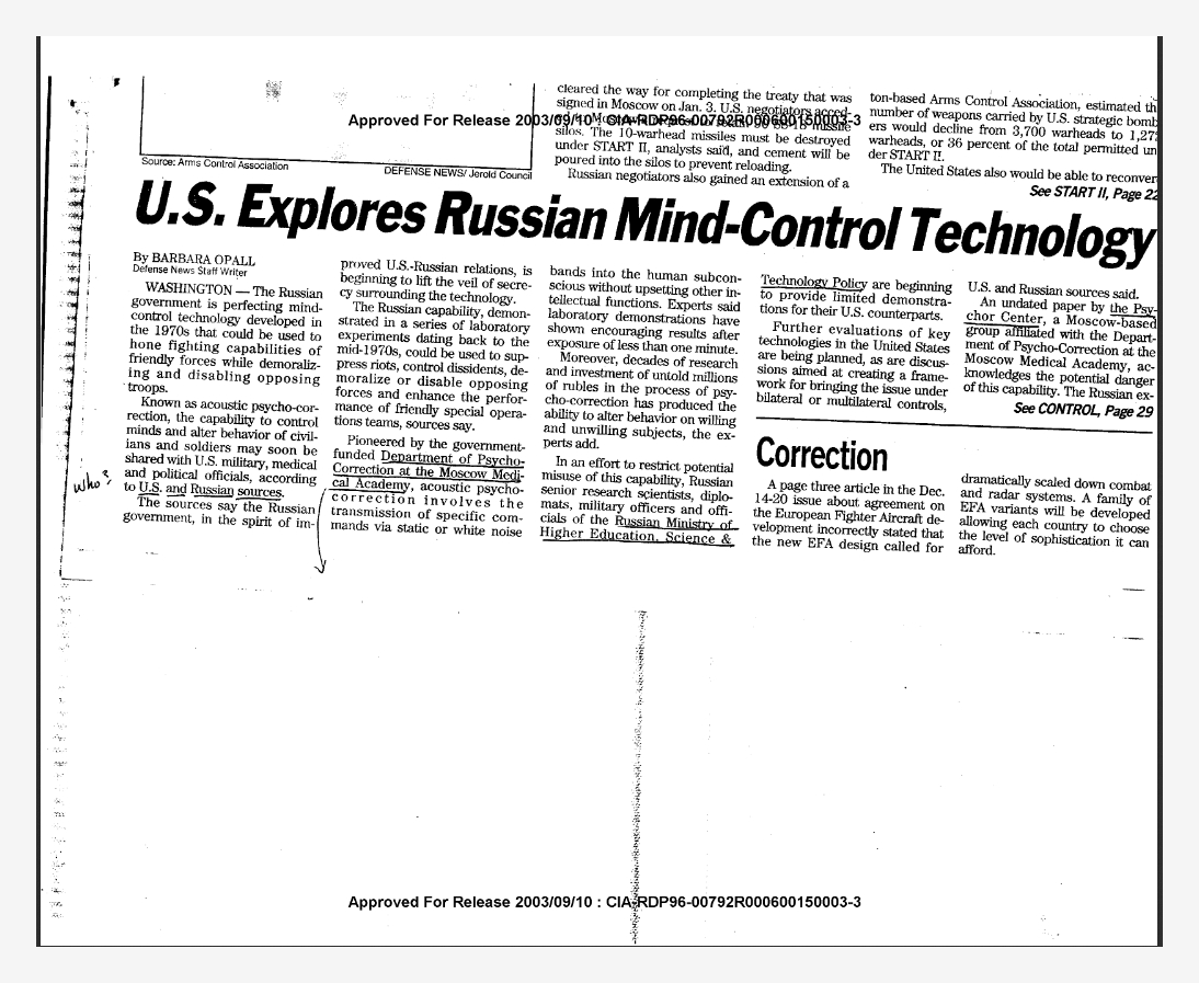
Les USA explorent la technologie russe de contrôle de l’esprit – janvier 1993

Ça fait pas mal de boulot, mais je vais commencer une série de traduction d’articles issus du site MindJustice.

Il y a une section qui traite d’articles extraordinaires « Sample of Rare and Outstanding Articles from 1976 to 1996 »

Le premier de la série est tiré de « Defense News 11-17 janvier 1993, p. 29 » dont on trouve la copie sur le site de la CIA, rien que ça.  https://www.cia.gov/library/readingroom/docs/CIA-RDP96-00792R000600150003-3.pdf


Les USA explorent la technologie russe de contrôle de l’esprit. Les États-Unis et la Russie espèrent protéger les techniques de contrôle de l’esprit par Barbara Opall, rédactrice de l’équipe (Defense News 11-17 janvier 1993, p. 29)


Washington – Le gouvernement russe perfectionne une technologie de contrôle de l’esprit mise au point dans les années 1970 qui pourrait être utilisée pour renforcer les capacités de combat des forces amies tout en démoralisant et en neutralisant les troupes adverses.

Connue sous le nom de psychocorrection acoustique, la capacité de contrôler les esprits et de modifier le comportement des civils et des soldats pourrait bientôt être partagée avec les autorités militaires, médicales et politiques américaines, selon des sources américaines et russes.

Les sources affirment que le gouvernement russe, dans un esprit d’amélioration des relations américano-russes, commence à lever le voile du secret entourant cette technologie.

La capacité russe, démontrée dans une série d’expériences en laboratoire remontant au milieu des années 1970, pourrait être utilisée pour réprimer les émeutes, contrôler les dissidents, démoraliser ou désactiver les forces adverses et améliorer la performance des équipes d’opérations spéciales amies, selon certaines sources.

Pionnière dans le domaine de la psychocorrection à l’Académie de médecine de Moscou, la psychocorrection acoustique, financée par le gouvernement, implique la transmission de commandes spécifiques par des bandes de bruit statique ou blanc dans le subconscient humain sans perturber les autres fonctions intellectuelles. Selon les experts, les démonstrations en laboratoire ont montré des résultats encourageants après une exposition de moins d’une minute.

En outre, des décennies de recherche et d’investissement d’innombrables millions de roubles dans le processus de psycho-correction a débouché sur la capacité de modifier le comportement des sujets volontaires et non volontaires, les experts ont précisé.

Dans un effort visant à limiter l’utilisation potentiellement abusive de cette capacité, des chercheurs scientifiques russes de haut niveau, des diplomates, des officiers militaires et des fonctionnaires du ministère russe de l’enseignement supérieur, des sciences et de la politique technologique commencent à fournir des démonstrations limitées pour leurs homologues américains.

D’autres évaluations des technologies clés aux États-Unis sont prévues, de même que des discussions visant à créer un cadre pour soumettre la question à des contrôles bilatéraux ou multilatéraux, selon des sources américaines et russes.

Un document non daté du Psychor Center, un groupe basé à Moscou et affilié au Département de psychocorrection de l’Académie de médecine de Moscou, reconnaît le potentiel ( ?) de cette capacité.

Les États-Unis et la Russie espèrent protéger les techniques de contrôle de l’esprit

Contrôle, à partir de la page 4

Les experts russes, dont George Kotov, un ancien général du KGB qui occupe actuellement un poste de ministre, présentent dans leur rapport une liste de logiciels et de matériel associés à leur programme de psycho-correction qui pourraient être achetés pour aussi peu que 80 000 $.

« Dans la mesure où il est devenu possible de sonder et de corriger le contenu psychique des êtres humains malgré leur volonté et leur conscience par des moyens instrumentaux, les résultats obtenus peuvent échapper à {notre} contrôle et être utilisés à des fins inhumaines de manipulation du psychisme », affirme le document.

Les auteurs russes notent que  » l’opinion mondiale n’est pas prête à traiter de manière appropriée les problèmes posés par la possibilité d’un accès direct à l’esprit humain « . C’est pourquoi les auteurs russes ont proposé la création d’un centre bilatéral pour les psychotechnologies où les États-Unis et le Russe {?} restreignent les capacités émergentes.

Janet Morris, du Global Strategy Council, un groupe de réflexion établi à Washington par Ray Cline, ancien directeur adjoint de la Central Intelligence Agency, est un agent de liaison clé entre les responsables russes et américains.

Dans une interview accordée le 15 décembre, Mme Morris a déclaré qu’elle et l’International Healthline Corp. de Richmond, en Virginie, avaient informé de hauts responsables du renseignement et de l’armée américaine des capacités russes, qui, selon Mme Morris, pourraient inclure des dispositifs portables pour des opérations spéciales, le contrôle des foules et des actions antipersonnel. Healthline Corp. évalue les technologies de soins de santé russes et financera des démonstrations russes aux États-Unis.

« Nous avons parlé de l’utiliser pour filtrer et préparer le personnel des opérations spéciales pour les missions extrêmement difficiles et des moyens de l’intégrer {?} pour les opérations psychologiques « , a dit M. Morris.

Elle a déclaré que les responsables de l’Armée de terre s’inquiétaient de la capacité des systèmes blindés et du personnel de l’Armée de terre d’utiliser les liaisons de communications électroniques. Les troupes au sol, dit-elle, risquent d’être exposées à des ondes sonores conductrices des os qui ne peuvent être compensées par des bouchons d’oreille ou d’autres équipements de protection actuels. M. Morris a ajouté que les contre-mesures américaines pourraient inclure l’annulation du son, un processus complexe qui consiste à diffuser des formes d’ondes à phases opposées dans des fréquences précisément adaptées.

Le major Pete Keating, porte-parole de l’armée américaine, a déclaré que les hauts responsables de l’armée américaine avaient exprimé leur intérêt pour l’examen des capacités russes, mais que Donald Atwood, secrétaire adjoint à la défense, avait rejeté les projets répétés de visites dans l’ancienne Union soviétique. Keating a dit qu’il ne connaissait pas la technologie de contrôle de l’esprit.

Selon des sources américaines, les responsables gouvernementaux et les dirigeants du monde des affaires et du monde médical examineront les offres de la Russie de placer la capacité de contrôle des esprits sous contrôle bilatéral.

Au moins un sénateur américain de haut rang, des responsables du renseignement du gouvernement et le Bureau des opérations, des plans et du développement des forces de l’armée américaine sont intéressés à examiner les capacités russes, ont indiqué des sources américaines.

En outre, International Healthline Corp. prévoit de faire venir une équipe de spécialistes russes dans les deux prochains mois pour démontrer cette capacité, a déclaré le président de la société, Jim Hovis, dans une interview accordée le 2 décembre.

Pendant ce temps, le Centre de recherche, de développement et d’ingénierie de l’armement de l’armée américaine mène une étude d’un an sur la technologie des faisceaux acoustiques qui pourrait refléter certains des effets rapportés par les Russes.

Le porte-parole de l’armée Bill Har ? a déclaré le 3 décembre que le commandement attendait l’étude d’un an commandée par Scientific Applications & Research Associates de Huntington Beach, Californie. Selon des sources américaines et russes, des recherches connexes menées à l’Institut Andreev, basé à Moscou, sont en cours.

Malgré l’intérêt croissant que suscite une capacité traditionnellement réservée aux romans de science-fiction, l’industrie et les experts universitaires sont prudents et sceptiques quant à son utilisation potentielle sur le champ de bataille.

« Ce n’est pas quelque chose qui me semble nécessiter une attention particulière « , a déclaré Raymond Gartho Gartho, analyste de la défense et du renseignement à la Brookings Institution de Washington, dans une interview accordée à De ?

Morris soutient que la capacité a été démontrée dans le laboratoire russe et qu’elle devrait être soumise à des restrictions internationales le plus tôt possible.

Catégories
Armes electroniques

Les armes V2K, manipulation des êtres humains.

https://www.wired.com/2008/05/army-removes-pa/


TRADUCTION

La très étrange page web de l’armée sur les armes « Voice-to-Skull » a été supprimée. C’était étrange qu’il soit là, et c’est encore plus étrange qu’il soit parti. Si vous le recherchez sur Google, vous verrez l’entrée pour « Voice-to-Skull device », mais, si vous cliquez sur le site, le lien est mort.

L’article, toujours disponible sur le site Web de la Federation of American Scientists, se lit comme suit :

Arme non létale qui comprend (1) un dispositif neuroélectromagnétique qui utilise la transmission par micro-ondes de sons dans le crâne de personnes ou d’animaux au moyen d’un rayonnement micro-ondes modulé par impulsions ; et (2) un dispositif sonore silencieux qui peut transmettre les sons dans le crâne de personnes ou d’animaux. REMARQUE : La modulation sonore peut être des messages vocaux ou audio subliminaux. Une des applications de V2K est l’épouvantail électronique pour effrayer les oiseaux à proximité des aéroports.

Le groupe britannique Christians Against Mental Slavery (Chrétiens contre l’esclavage mental) a noté le changement pour la première fois (ils ont aussi une copie d’écran permanente de la page). Un représentant du groupe me dit qu’ils ont contacté le webmestre, qui leur a seulement dit que l’entrée était « définitivement supprimée ».

L’image ci-dessus est une représentation autoproclamée par une personne de la façon dont une arme « voix vers crâne » pourrait fonctionner.


The Army’s very strange webpage on « Voice-to-Skull » weapons has been removed. It was strange it was there, and it’s even stranger it’s gone. If you Google it, you’ll see the entry for « Voice-to-Skull device, » but, if you click on the website, the link is dead.

The entry, still available on the Federation of American Scientists‘ website reads:

Nonlethal weapon which includes (1) a neuro-electromagnetic device which uses microwave transmission of sound into the skull of persons or animals by way of pulse-modulated microwave radiation; and (2) a silent sound device which can transmit sound into the skull of person or animals. NOTE: The sound modulation may be voice or audio subliminal messages. One application of V2K is use as an electronic scarecrow to frighten birds in the vicinity of airports.

The U.K.-based group Christians Against Mental Slavery first noted the change (they also have a permanent screenshot of the page). A representative of the group tells me they contacted the Webmaster, who would only tell them the entry was « permanently removed. »

The image above is one person’s self-styled depiction of how a « voice-to-skull » weapon might work.

[Image: Raven1.net]

ALSO:


Catégories
Armes electroniques

Clubic : Un laser capable de vous envoyer un son sans que personne d’autre ne l’entende.

Des chercheurs du MIT ont mis au point un dispositif permettant d’envoyer un son directement à l’oreille d’une personne. Reposant sur un système de laser, il est capable de transmettre précisément un signal audio, de sorte que seul le destinataire puisse l’entendre.

Et si on pouvait transmettre les sons aussi précisément que la lumière via un laser ? C’est le résultat qu’ont obtenu des chercheurs du Laboratoire Lincoln, du MIT (États-Unis).

Lumière + vapeur d’eau = son

Les scientifiques se sont en effet inspirés du fonctionnement du laser. Leur système repose sur la photoacoustique, c’est-à-dire la génération d’ondes sonores à partir d’interactions entre la lumière et la matière. En l’occurrence, c’est la vapeur d’eau contenue dans l’air qui réagit avec l’émetteur laser élaboré par les chercheurs, pour produire le signal audio.

De plus, les auteurs de l’étude ont réussi à exploiter une longueur d’onde fortement absorbée par l’eau, ce qui permet d’augmenter la puissance et la qualité du son produit. Par conséquent, leur dispositif peut fonctionner même dans un environnement relativement sec, ne nécessitant qu’une quantité restreinte de vapeur d’eau.

Un « laser sonore » précis et sûr

Le système ainsi créé permet donc de générer un flux audio et de le transmettre de façon précise jusqu’à son destinataire, et uniquement à celui-ci, même dans un environnement bruyant. Et ce, sans risque pour les yeux ni la peau.

Les tests réalisés ont prouvé son efficacité à une distance de 2,5 m, ce qui reste encore assez limité. Mais d’après les scientifiques, ce résultat n’a été obtenu qu’avec du matériel disponible dans le commerce, et cette performance pourrait donc être aisément améliorée. Ce qui offrirait la possibilité de chuchoter à l’oreille d’une personne, à plusieurs mètres de distance.

Source : BGR


Toujours présenté pour une bonne cause, en omettant les graves dérives possibles. Il n’ échappera bien évidement qu’à l’auteur de l’article, que ceci rend obsolète les diagnostiques psychiatriques concernant la schizophrénie. Tellement insignifiant qu’il est absolument inutile de le mentionner… De même, est il bien nécessaire de parler de voice to skull, la « voix de Dieu » ?

WASHINGTON — Researchers have demonstrated that a laser can transmit an audible message to a person without any type of receiver equipment. The ability to send highly targeted audio signals over the air could be used to communicate across noisy rooms or warn individuals of a dangerous situation such as an active shooter.


New Technology Uses Lasers to Transmit Audible Messages to Specific People

Photoacoustic communication approach could send warning messages through the air without requiring a receiving device

WASHINGTON — Researchers have demonstrated that a laser can transmit an audible message to a person without any type of receiver equipment. The ability to send highly targeted audio signals over the air could be used to communicate across noisy rooms or warn individuals of a dangerous situation such as an active shooter.


In The Optical Society (OSA) journal Optics Letters, researchers from the Massachusetts Institute of Technology’s Lincoln Laboratory report using two different laser-based methods to transmit various tones, music and recorded speech at a conversational volume.

“Our system can be used from some distance away to beam information directly to someone’s ear,” said research team leader Charles M. Wynn. “It is the first system that uses lasers that are fully safe for the eyes and skin to localize an audible signal to a particular person in any setting.”

Creating sound from air

The new approaches are based on the photoacoustic effect, which occurs when a material forms sound waves after absorbing light. In this case, the researchers used water vapor in the air to absorb light and create sound.

“This can work even in relatively dry conditions because there is almost always a little water in the air, especially around people,” said Wynn. “We found that we don’t need a lot of water if we use a laser wavelength that is very strongly absorbed by water. This was key because the stronger absorption leads to more sound.”

One of the new sound transmission methods grew from a technique called dynamic photoacoustic spectroscopy (DPAS), which the researchers previously developed for chemical detection. In the earlier work, they discovered that scanning, or sweeping, a laser beam at the speed of sound could improve chemical detection.

“The speed of sound is a very special speed at which to work,” said Ryan M. Sullenberger, first author of the paper. “In this new paper, we show that sweeping a laser beam at the speed of sound at a wavelength absorbed by water can be used as an efficient way to create sound.”

Laboratory tests

In the lab, the researchers showed that commercially available equipment could transmit sound to a person more than 2.5 meters away at 60 decibels using the laser sweeping technique. They believe that the system could be easily scaled up to longer distances. They also tested a traditional photoacoustic method that doesn’t require sweeping the laser and encodes the audio message by modulating the power of the laser beam.

“There are tradeoffs between the two techniques,” said Sullenberger. “The traditional photoacoustics method provides sound with higher fidelity, whereas the laser sweeping provides sound with louder audio.”

Next, the researchers plan to demonstrate the methods outdoors at longer ranges. “We hope that this will eventually become a commercial technology,” said Sullenberger. “There are a lot of exciting possibilities, and we want to develop the communication technology in ways that are useful.”

Paper: R. M. Sullenberger, S. Kaushik, C. M. Wynn. “Photoacoustic communications: delivering audible signals via absorption of light by atmospheric H2O,” Opt. Lett., 44, 3, 622-625 (2019).
DOI: https://doi.org/10.1364/OL.44.000622.

About Optics Letters
Optics Letters offers rapid dissemination of new results in all areas of optics with short, original, peer-reviewed communications. Optics Letters covers the latest research in optical science, including optical measurements, optical components and devices, atmospheric optics, biomedical optics, Fourier optics, integrated optics, optical processing, optoelectronics, lasers, nonlinear optics, optical storage and holography, optical coherence, polarization, quantum electronics, ultrafast optical phenomena, photonic crystals and fiber optics.

About The Optical Society

Founded in 1916, The Optical Society (OSA) is the leading professional organization for scientists, engineers, students and business leaders who fuel discoveries, shape real-life applications and accelerate achievements in the science of light. Through world-renowned publications, meetings and membership initiatives, OSA provides quality research, inspired interactions and dedicated resources for its extensive global network of optics and photonics experts. For more information, visit osa.org.

Catégories
Armes electroniques General

Gouverner par le chaos (chapitre: Le Mind Control)

Gouverner par le chaos

Ingénierie sociale et mondialisation

Table des matières

Politique et massification
Politique et mondialisation
Qu’est-ce que l’ingénierie sociale?
La stratégie du choc
La conduite du changement
Le social learning
La fabrication du consentement
Le tittytainment
Le pied-dans-la-porte
Le mind control
Le virtualisme
La guerre contre-insurrectionnelle
Le reality-building
Le management négatif
La théorie de la Jeune-Fille
Le biopouvoir
Conclusion provisoire
L’Appel des Résistants


Le Mind Control

Faisons encore un pas dans la régression mentale
provoquée et le hacking psychosocial. Chacun se souvient
des propos de Patrick Le Lay, alors Président Directeur
Général de TF1, sur « le temps de cerveau disponible »
que sa chaîne de télévision vendait aux annonceurs
publicitaires. Rien d’anecdotique dans cette formulation.
Après le contrôle des émotions et des situations,
l’ingénierie sociale s’est beaucoup intéressée au contrôle
direct du cerveau, dans l’optique de court-circuiter le
champ des représentations pour s’attaquer directement à
la programmation du système nerveux dans sa
matérialité la plus brute. Cette analogie entre cerveau et
ordinateur, déjà perceptible dans la cybernétique, le
cognitivisme et le Social Learning, s’appuie en fait sur le
Learning tout court, c’est-à-dire les théories de
l’apprentissage, au sens de « apprendre à un être vivant à
se comporter de telle façon ». Pour le dire frontalement,
le Learning est la science du dressage et duconditionnement
comportemental.Elle fut originellement testée sur des animaux de laboratoire,
mais rapidement appliquée à l’humain dès les années
1940 au travers des recherches en Mind Control (contrôle
mental), ou MK (Mind Kontrolle), menées dans le but de
créer des Candidats Mandchous et des soldats parfaits,
ignorant la peur, insensibles à la douleur, etc. Divers
protocoles furent mis au point, s’appuyant sur lesprincipes behaviouristes de « conditionnement classique
», issus des travaux de Pavlov sur les réflexes
conditionnés (stratégie directe et déterministe) et de «
conditionnement opérant », issus des travaux de Skinner
sur l’induction de comportements à partir du façonnage
de l’environnement (stratégie indirecte et tendancielle).
Le jeu sur la récompense et la punition pouvant aller
jusqu’à des actes de torture, on ne s’étonnera pas que le
programme de recherche MK-Ultra, dont les dossiers ont
été récemment déclassifiés par la CIA après avoir été top-
secrets pendant une cinquantaine d’années, ait fortement
inspiré non seulement l’ouvrage déjà mentionné de
Naomi Klein mais encore l’enquête très approfondie de
Gordon Thomas, intitulée Les armes secrètes de la CIA —
Tortures, manipulations et armes chimiques. L’auteur y
restitue l’historique complet du projet MK-Ultra, avec ses
savants fous affairés autour de leurs cobayes humains, ou
« sujets jetables », expliquant que la germanisation du
terme control en Kontrolle était un clin d’œil aux origines
des scientifiques qui développèrent les premiers ces
recherches, d’anciens nazis exfiltrés après la guerre aux
États-Unis ou en Angleterre dans le cadre de l’opération
Paperclip. Ainsi, depuis 1945 et dans la continuité de ce
que les scientifiques du Troisième Reich avaient
commencé de mettre au point, de nombreuses
expériences sur l’hypnose, les hallucinogènes, l’influence
subliminale, le lavage de cerveaux et la re-
programmation mentale furent (et continuent d’être)
élaborées sur les individus et sur les masses à l’Institut
Tavistock, au Massachusetts Institute of Technology
(MIT), ou sur d’autres campus universitaires tels que
Harvard. Le malheureuxTed Kaczynski, devenu célèbre sous le pseudonyme «
Unabomber », en fut lui-même victime au début des
années 60, alors qu’il était encore étudiant sous la
direction de Henry A. Murray. Plus récemment, on a vu
naître de ces recherches une nouvelle discipline, le
neuromarketing, fondée sur l’imagerie médicale du
cerveau et visant explicitement à déclencher des pulsions
d’achat irrépressibles par l’activation ciblée de certaines
zones du système nerveux.
Le Mind Control est friand de métaphores
informatiques et relatives à l’Intelligence Artificielle, son
projet consistant à réécrire le programme
comportemental d’une machine vivante mais sans que
cette machine ne s’en rende compte. Piratage psycho-
socio-biologique, où le code source du sujet cobaye a été
craqué, puis effacé et reformaté par une entité extérieure
au sujet, qui s’est ainsi rendue propriétaire de
l’inconscient du sujet et qui peut donc orienter son
devenir. Un hacker s’est infiltré dans la mémoire, en a
pris le contrôle, l’a reconfigurée selon ses plans, a
implémenté de nouveaux habitus, de nouveaux
algorithmes comportementaux et pilote désormais la
machine humaine à distance. Mais surtout, il a effacé
toute trace de son effraction et de sa manipulation. La
philosophie du Mind Control, l’emprise totale sur un être
vivant, emprise autorisée par la réduction de cet être à
une machine computationnelle simplement animée
d’entrées et de sorties d’information (input et output), a
ainsi infusé toute la politique moderne, progressivement
réduite à la gestion de flux quantitatifs. La cybernétique,
même quand elle se veut « humaniste » dans les
conférences de la Fondation Macy (1946-1953) ou dans lerapport Meadows du Club de Rome (1972), ne peut
s’empêcher de chercher à réduire l’incertitude à zéro et
donc à produire un effet de « chosification » du vivant.
Ces diverses approches de la gestion des groupes
humains ont toutes en commun de produire des effets de
nivellement par le bas. À chaque fois, il s’agit de
contourner le lobe frontal du cerveau, le néocortex, siège
du langage et des fonctions dialectiques, pour prendre
directement le contrôle des fonctions pré-linguistiques :
les réflexes primitifs du cerveau reptilien, et les émotions
dans le système limbique. Il s’agit de rendre impossible la
sublimation, c’est-à-dire de désirer des mots plutôt que
des objets, et de maintenir toute la vie entre deux états
mentaux simplifiés pré-langagiers, dérivés des deux
émotions primitives que sont la peur et l’excitation
érogène. Cette atrophie du champ psychique génère
évidemment toute une gamme d’états dépressifs et de
pathologies mentales diverses, que l’on peut rassembler
sous les termes de désymbolisation, de perte de Sens et
de structure mentale. Mais pour parvenir à ses fins, à
savoir la construction d’un système social totalement sûr
et prévisible, l’ingénierie politique des pays développés
n’a pas eu d’autre choix que de considérer l’humain
comme moins qu’un animal : comme un simple objet
plastique et à disposition pour le recomposer à loisir.

Catégories
Armes electroniques

MICROWAVE BIOEFFECT CONGRUENCE WITH SCHIZOPHRENIA

MICROWAVE BIOEFFECT CONGRUENCE WITH SCHIZOPHRENIA


John J. McMurtrey M. S.,[a] Copyright 2002, 10 Apr. 2005

Co-authorship is negotiable towards professional publication in an NLM indexed journal, Email- Johnmcmurt@aol.com

Donations toward future research are gratefully appreciated at http://www.slavery.org.uk/FutureResearch.htm


ABSTRACT

The substantiation for microwave voice transmission development, which can be isolated to an individual, prompts review of the correlation between microwave bioeffects and schizophrenia. These correlations are extensive. Studies of both conditions report short-term and spatial memory deficit, time estimation changes, deficits in sequencing, coordination deficit, numerous electrophysiologic changes, startle decrease, neurotransmitter changes, hormone alterations, immune alterations, mitochondria deficits, lipid phosphorylation decrease, lipid peroxidation, deleterious histologic change in disease reduced brain areas, activation of hallucination involved brain areas, and ocular disease. Schizophrenia findings correlate with microwave bioeffects so extensively as to indicate a congruence, and appear to implicate a microwave involvement with enough patients to be remarkable in study results. The development of methods to exclude microwave means in psychosis is imperative, and research is proposed.


INTRODUCTION


Remote microwave induced sound [1] [2] [3] and internal voice technology has long been discovered, [4] developed, [5] [6] detailed in patents, [7] [8] [9] [10] with weapons applications described. [11] [12] [13] [14] That such technology can be applied remotely and coupled to target tracking technology [15] has implications for patients who, by virtue of voice transmission complaint and other symptoms, are diagnosed with various mental disorders. [16] Auditory hallucination is most prevalent in schizophrenia, which features in 60% of cases. [17] [18] A frequent patient understanding of the origin of voices is by remote transmission, though the very concept is considered delusional, [19] and often the diagnosis is psychosis of varying severity depending on functional ability, [20] without any investigation of described internal voice capabilities.

The substantiation of microwave voice transmission development suggests examination of any microwave bioeffect correlation with schizophrenia findings.  The hypothesis tested was that perhaps some discrepant schizophrenia study results could differentiate patients subjected to technological assault. Unfortunately, little differentiation was evident, because the correlations appear too extensive, as presented in overview Table I. Unless otherwise noted, the microwave exposure effects examined are at low intensity, and are expressed in text parenthesis in terms of existing exposure standards. [21] [b] Since most of the observed correlations are close to microwave exposure standards, the possibility of an environmental microwave association with schizophrenia is considered.


Cognitive Function

Schizophrenics are particularly impaired in memory. [22], [23] Pulsed radar exposed Latvian children are deficient in short term memory compared to unexposed children. [24]   Rats exposed to microwaves during gestation exhibit conditioned avoidance acquisition deficit as adults (at 1.61 X US occup. std.). [25] Adult rat microwave exposure yields avoidance conditioning deficits (at 31% of US occup. std. & 1.75 X ICNIRP [c] pop. std.),[26] [27] and there is some mention of ‘retrograde amnesia’ with such conditioning (at 63% of US pop. std.). [28] [29] Schizophrenic ‘working memory’ is considered central to many schizophrenic symptoms. [30] Schizophrenia deficits are in multiple areas of working memory, and the disorder exhibits deficits specifically in spatial working memory. [31] [32] [33] [34] Rat spatial ‘working memory’ on microwave exposure is deficient for performance in a water maze, (1.2 X US pop. std.) [35] [36] and in the 12 arm radial maze (60% of US pop. std.), [37] [38] but apparently not when distal cues are present for radial mazes, [39] [40] which are preferred in rodents. [41] [42]

Schizophrenia time estimation is altered with overestimation of short time intervals. [43] [44] [45] [46] Microwave exposed rats, when trained on inter-response time reinforcements reflect the same direction of deficit by increased total lever presses (at 31% of US occup. std. & 1.75 X ICNIRP pop. std.), 25 26 shorter inter-response times (62% of US occup. std.), [47] which are even greater for pulsed microwaves (1.1 X US occup. std.). [48] A rat time estimation task on microwave pulsation indicated change in discriminating stimulus duration, increased time to complete tasks, and increased the amount of non-response (at 90% of ICNIRP pop. std.). [49] The authors suggest an effect on the animal’s internal clock.

Schizophrenia patients exhibit deficits in memory for temporal order. [50] [51] [52] [53] Microwave exposed rats with simple response sequence conditioning (½ to 1.6 X US occup. std.) [54] [55] [56] [57] [58] exhibit analogous sequencing deficits. In humans, the order threshold of discriminating the ear of first tone presentation as succeeded by a tone separated by decreasing intervals to the other ear, increases after 50 minutes cell phone exposure, while the threshold decreases with no exposure rest. [59]

The hippocampus has general importance to memory, [60] while sub serving spatial, temporal, and sequence memory. [61] In rodents the hippocampus is one of the most responsive brain regions to microwave exposure (at US pop. std. to ½ US occup. std.), [62] [63] and microwave induced histologic damage is observed (at ¼ ICNIRP pop. std. to 1.8 X US occup. std.). [64] [65] [66] Some schizophrenics have anti-hippocampal antibodies, [67] and


Table I


SCHIZOPHRENIA SIGN/SYMPTOM CORRELATION WITH MICROWAVE BIOEFFECTS


  Cognitive/Physiologic Parameter


Schizophrenia Sign/Symptom Microwave Bioeffect


Cognitive Function


         Memory Deficit Deficits in Memory and Working Memory Child Short Term Memory Deficit, Rat Conditioned Avoidance and Spatial Memory Deficits
         Time Estimation Overestimation of Short Intervals Rat Shortened Inter-Response Times and Increased Responses in Time Estimation Tasks
         Temporal Order Temporal Order Memory Deficits Human Decrease in Temporal Order Discrimination, Rat Response Sequencing Deficits
  Startle Response Decreased in Some Patients Decreased in Animals
  Coordination/Balance Decreased Coordination and Balance Decreased Child Coordination, Rat Decrease in Coordination and/or Balance
   

Electrophysiology


  Contingent Negative Variation Decreased in Patients Decreased on Human Cell Phone Exposure
  Event Related Auditory Response Decreased in Patients Decreased in Animals, Component Decrease in Human Cell Phone Exposure
  EEG Delta Waves Increased in Patients Increased in Humans and Animals
  EEG Beta Waves Increased in Patients Increased in Humans and Rats


TABLE I, continued


SCHIZOPHRENIA SIGN/SYMPTOM CORRELATION WITH MICROWAVE BIOEFFECTS


Physiologic Parameter Schizophrenia Sign/Symptom


Microwave Bioeffect
 

Neurotransmitters


       Dopamine Indicated Decreased in Negative Symptom Schizophrenia Indicated Decreased Based on Extensive Evidence
       Serotonin Indicated Decreased in Patients Based on Numerous Studies Found Decreased in Rats
    γ-Aminobutyric Acid Decreased Uptake & Release in Schizophrenia Synaptosomes Decreased Receptor Specific Binding
    Acetylcholine α 7-nicotinic Receptor Decrease in Some Brain Areas Consistent with Acetylcholine Decrease Decreased Rat Acetylcholine Release, and Precursor Uptake in Same Brain Areas
Hormones


       Corticosteroids ACTH, Cortisol, and Corticosterone Reported Increased Adrenal Depletion with ACTH, Cortisol, and Corticosterone Increase Reported
       Melatonin Decrease Reported in Some Patients Decreased on Human Cell Phone and EMF Exposure
Mitochondria Decreased Number in Schizophrenia Brain Deleterious Changes with Decreased ATP, Creatine Phosphate, and Marker Enzymes
Immunology


       Autoimmunity Suggested from Autoantibody Levels and Autoimmune Disease Incidence Reported Induced and Stimulated
       Tumor Necrosis Factor Reported Increased Numerous Reports of Increase in Animals
       B Lymphocytes Balance of Evidence Shows Increase in Some Patients Increased in Mouse Spleen with Genetic Control


Table I, continued.


SCHIZOPHRENIA SIGN/SYMPTOM CORRELATION WITH MICROWAVE BIOEFFECTS


Physiologic Parameter Schizophrenia Sign/Symptom


Microwave Bioeffect
 

Lipids


       Phosphorylation Decreased on Magnetic Resonance Spectroscopy Decreased P32 Lipid Incorporation
       Peroxidation Increase in Patients Increased in vitro and in Rats
Blood-Brain Barrier Suggested Impaired in Patients Reported Decreased in Numerous Studies
 

Anatomy & Histology


       Hippocampus Hippocampal-amagdala Complex Volume Reduced

in Most Studies

Degenerative Hippocampus Histology Reported
       Thalamus Volume Reduction Observed in Many Studies Degenerative Histology Reported
       Cerebellum Changes Observed in Many Studies Degenerative Histology

Reported

       Cortex Volume Reductions Observed in Frontal and Parietal Cortex by Many Studies Several Reports of Degenerative Unspecified Cortex Histology
Metabolic Activation Hallucination Activates Temporal Lobe, and Thalamus with Collicular Activation Found in Some Studies Animal Activation of Temporal Lobe, Thalamus, and Inferior Colliculus on Hearing Effect Pulsed Microwaves
Ocular Disease


       Cataract Subcapsular Cataract Reported Without Association to Medication Known Cause of Subcapsular Cataract
       Retinopathy Associated with Widely Prescribed Anti-Psychotics Associated with Occupational Exposure and Experimentally Produced
Voice Transmission Hallucination Most Common Symptom Voice Transmission Affirmed


the same hippocampus CA1 region that is volume decreased in schizophrenia, [68] on microwave exposure shows altered neuronal activity in vitro slices from rats, [69] as well as decreased acetylcholine release in vivo rats (1/2 US occup. std.). [70] Mouse hippocampus mitochondrial activity is indicated decreased on microwave exposure (1/4 US pop. std.).[71] Although not actually affecting performance, cell phones are reported to affect a magnetoencephalographic (MEG) component of verbal memory encoding, suggesting interference. [72] Multiple human case reports of memory difficulty, with other neurasthenic complaints exist on excess microwave exposure. [73] [74] [75] Microwave exposed rats with avoidance conditioning, exhibit changes in emotion and integrative function [76] from which parallels to schizophrenia can be drawn.  Accidental and/or occupational 1-10 GHz excess radar exposure exhibits frontal lobe neuropsychiatric symptoms. [77]


Startle Response

Some schizophrenics have little or no startle response. [78] Microwave exposed rats exhibit decreased startle under both continuous wave [79] and pulsed [80] [81] conditions (1.2 X US occup. std.) with the latter decreasing startle in mice. [82]   Pre-natal rat exposure decreases startle in females (1.2 X US occup. std.). [83] Some schizophrenics are hypo- or non-responders to orienting responses [84] and normally evoked electrodermal activity. [85] Microwave occupational exposure inhibits galvanic skin response. [86] Rats also fight less on microwave exposure (6% & 23% of US pop. std.), [87] [88] will avoid hearing effect pulsed microwaves, [89] and mice decrease exposure by their orientation in a field. [90]


Coordination, Balance, and Exercise Tolerance

Schizophrenics have decreased ability in coordination tasks, and more instability in balance. [91] [92] [93] [94] Latvian children exposed to pulsed radar have less motor competence than unexposed children. 24 Microwave exposed rats show degradation of motor coordination and/or balance (at 21% of US pop. std.). [95]

High peak power pulsed microwave 25 minute exposures decreased rat treadmill running by about one-third. [96] A German abstract states schizophrenics could only achieve one-third of the aerobic-anaerobic threshold for untrained controls. [97] Schizophrenics have shown abnormal thermoregulation on exercise with greater increases in core temperature. [98] [99]


Electrophysiology

An electrophysiologic indicator of ‘working memory’, contingent negative variation (CNV) [100] is decreased in schizophrenia, [101] [102] [103] which is reported to correlate to ratings for negative symptoms of affective flattening and avolition-apathy. [104] Cell phone radiation also decreases human CNV. [105] [106] The test involves a warning stimulus and an imperative stimulus with the intervening evoked waveform representative of sensory and motor adjustment prior to expected action.

Electrophysiologic auditory event related P300 and antecedents are reduced in some schizophrenics, [107] [108] with increased latency indicated. [109] [110]   Decreased auditory event response is observed during hallucination in magnetoencephalographic (MEG) [111] and functional magnetic resonance imaging [112] studies, which resembles the interfering sound response. [113] Like hallucination or outside sound, microwave hearing exposure decreases cortical auditory evoked potential amplitudes with increased latency in rats, rabbits, (less than US occup. std.) [114] and cats. [115]   Schizophrenia auditory P300 reduction is related to deleterious signs and poor prognosis. [116] The human N100 amplitude is decreased on GSM cell phone exposure, [117] [118] which is also decreased in schizophrenia [119] [120] [121] with the reduction correlating to withdrawal-retardation scores, [122] and paranoid diagnosis. [123]

Hearing effect pulsed microwaves evoke brain responses similar to auditory stimuli. [124] [125] [126]  Radio frequency exposure increases human hearing threshold for auditory tones. [127] Sound also decreases the brain stem microwave hearing response. [128]

Auditory brain stem responses (ABR) in schizophrenics having hallucination, [129] [130] [131] never medicated hospitalization, [132] marked personality deterioration, [133] and negative symptoms [134] involve abnormalities of increased peak latency and missing peaks. Since microwave hearing produces an ABR, [135] [136] interference is expected, which would complicate ABR topographic appearance. Increased ABR latency is reported from a cell phone study, [137] though this is not replicated by all cell phone studies. [138] [139]

Soviet and American microwave exposure of humans report EEG increases in delta or “slow” waves, abnormal to adult alertness in quantity. Acute human exposure to continuous or pulsed microwaves, exhibit increased electroencephalogram (EEG) delta waves (less than US pop. std.). [140] Soviet and East European microwave occupational exposure review observes increased EEG delta waves. [141] Cell phones also increase human delta waves in adults [142] and children. [143]

Rabbit and rat microwave irradiation yield delta waves as well. Daily 3 hour rabbit exposures produces delta wave increases at 1 month to pulsed microwaves and at 2 months to continuous wave exposure (1/2 US occup. std.). [144] Daily 7 hours of microwave exposure produced delta waves after 10-15 days in rabbits at 1/3rd the US population exposure standard, but took 1 month for delta wave increase at 1/30th this standard. [145] Rat microwave irradiation induces delta waves in the left hemisphere by continuous wave, but in the right hemisphere when modulated. [146] Delta waves are also produced by extra low frequency radiation in rabbits [147] or magnetic fields in humans. [148]

Microwave delta wave increases correspond to delta wave increases widely noted in untreated, [149] [150] [151] [152] [153] [154] [155] [156] [157] [158] [159] [160] [161] and medicated [162] [163] [164] [165] [166] [167] [168] [169] schizophrenia EEGs. [170] Delta waves particularly correspond to psychotic episodes, [171] [172] and occur immediately prior to auditory hallucination. [173]   Higher delta power correlates with negative schizophrenia symptoms, [174] [175] and ‘psychomotor poverty’, [176] [177] while higher left temporal delta wave dipole density correlates to ratings for hallucination and paranoia. [178]

Intermittent long-term occupational exposure to microwaves increases EEG beta frequencies. [179] A therapeutic microwave instrument immediately increased beta wave power in humans, and cell phones increase these frequencies after a 15 minute delay. 142  Cell phones also increase human beta waves during tasks. [180] Microwave exposure increases beta frequencies in the rat (at ½ pop. std. to 1.2 X occup. std.). [181] [182] [183]

Though some anti-psychotic drugs decrease beta frequencies, schizophrenia EEG studies exhibit increased beta frequencies. 150 151 152 154 155 156 160 161 162 167 175 [184] [185] [186] [187] [188] [189] [190] MEG frequencies in the beta band are observed on auditory hallucination. [191] Treatment-resistant patients have greater increases in beta frequencies above 18 Hz, [192] [193] with dipole location sources of beta frequencies varying on auditory stimulation according to symptom severity. [194] Greater increase in beta frequencies is associated with decreased mismatch negativity amplitudes, [195] and ‘psychomotor poverty’. 174 176 177  The sources of increased schizophrenia beta frequencies are also more anterior and superficial than controls. [196] [197] Superficial tissue absorbs more microwave energy than deep tissues. [198]

Electromagnetic field EEG entrainment occurs especially within physiologic brain frequencies (1-40 Hz.), either with a so modulated carrier wave or at these extra low frequencies. Microwave EEG entrainment (or change to exposure frequency) is demonstrated in cats, [199] and rats. [200] Lower frequency radiation or magnetic EEG entrainment is observed in rabbits, [201] monkeys, [202] and humans. [203] In addition to the capacity of entrainment to produce delta or beta waves, the effect forms a basis for schizophrenic thought interference complaints, and is of non-lethal weapon concern. [204]


Neurotransmitters

Both schizophrenia and microwave exposure involve brain dopamine alterations. Many have long attributed positive schizophrenic symptoms to dopamine increases based on differential drug effects. [205] However, findings in schizophrenics with negative symptoms for dopamine metabolites, dopamine receptors, and drug studies indicate decreased dopamine. [206] Based on behavioral changes, drug study results, and enzyme alterations, microwave exposure also indicates decreased dopamine. [207] [208] [209]

Other neurotransmitter alterations correspond in both microwave bioeffects and schizophrenia. Brain postmortem tissue analysis, cerebrospinal fluid, and drug studies find decreased schizophrenia serotonin. [210] Although rat serotonin metabolite ratios indicate increased serotonin turnover rates on acute microwave exposure (3.1 X US pop. std.), [211] brain serotonin decrease occurs on prolonged exposure (near US occup. std.). 126 Rat microwave exposure from birth to 15 days decreased serotonin in adults (near ½ US occup. std.). [212]

Cortical synaptosome γ-aminobutyric acid (GABA) uptake and release is reported decreased in schizophrenics, who have decreased GABA neurons, [213] and synthetic enzymes. [214]   GABA receptor binding (by 3H-muscimol) decreases in rat neocortex on microwave irradiation (2.6 X US occup. std.). [215] Immunohistochemistry also indicates decreases in rat cellular GABA content in Purkinje cells of the cerebellum (10 X ICNIRP occup. std.). [216]

There is evidence for a cholinergic decrease in Lewy Body Syndrome, which is a psychosis that can have schizophrenia diagnosis, [217] and there is consistent evidence for a decrease in the α 7-nicotinic acetylcholine receptor in schizophrenia hippocampal and frontal areas, [218] which indicates decreased acetylcholine levels. [219] Acetylcholine release is found decreased on in vivo rat microwave exposure for the hippocampus (1/2 US occup. std.). 70 Acute rat microwave exposure also decreases sodium dependent choline uptake, the rate limiting step in acetylcholine synthesis, especially in frontal cortex followed by the striatum on either pulsed or continuous wave, but only pulsation decreased hippocampal choline uptake (60 % of US pop. std.). 37 [220] [221] The hippocampus and striatum are limbic structures– a brain system prominent in schizophrenia pathogenesis, which is implicated in microwave bioeffects, [222] and rats differently responsive to the vocalizations of other shocked rats, differ in behavior and neurotransmitter levels on very low microwave exposure (1/2 % of US pop. std.). [223]


Hormones

Corticotrophin is indicated to mediate microwave stress, [224] [225] and microwaves influence adrenal steroids. Satellite station operator microwave exposures produce a stress reaction of urinary increases in 11-oxycorticosteroids and stress hormone diurnal pattern shift (1/10th of US pop. std.). [226] Cell phone exposure transiently increases blood cortisol levels. [227] Rat microwave exposure yields adrenal activation resulting in adrenal medulla epinephrine and corticosteroid depletion (1.8 X US occup. std.). [228] Female rat microwave exposure increased blood corticosterone and ACTH, with decreased estradiol independent of pregnancy (1.2 X US pop. std. to 1.2 X US occup. std.). [229] [230] [231] Schizophrenic patients have increased cortisol [232] with less dexamethasone cortisol suppression than controls, [233] [234] and corticosterone increase is reported. [235] Schizophrenics have such hypothalamic-pituitary-adrenal axis over activity with ACTH increase as to feature the metabolic syndrome. [236] Patient cortisol lacks sleep inhibition, and correlates with paranoia and hallucination.

Decreased melatonin is consistently reported in schizophrenia, [237] [238] [239] [240] [241] with such a finding in paranoid patients. [242] [243] Electromagnetic fields diminish melatonin in animals. 207 [244] Human melatonin decrease is both at lower frequency exposure, [245] [246] [247] [248] and on cell phone use. [249] The pineal gland synthesizes melatonin from serotonin, [250] also decreased as above. Abnormal EEG and decreased melatonin are associated with pineal calcification, [251] which has lower incidence in undeveloped societies [252] who also show better schizophrenic prognosis. [253]


Mitochondria Changes

Mitochondria are altered in both schizophrenia and microwave exposure. Mitochondria deformation, size reduction, and decrease in number from 20-33% in schizophrenia brain are observed. [254] Cytochrome c oxidase, of the mitochondria oxidative phosphorylation system, is reduced from 30-63% in the schizophrenic brain. [255] Schizophrenic mitochondria gene expression is decreased in five pathways. [256] Acute microwave exposure evidences mitochondria matrix density decrease, and cristae degeneration in vitro for liver cells (1.2 X US occup. std.), [257] with pulsation experiments inducing normal cristae pattern loss, lamellar body formation, and mitochondrial membrane breaks in neuroblastoma cells. [258] Adenosine triphosphate (ATP) and creatine phosphate (CP) levels depend on oxidative phosphorylation, which requires electron transport components of mitochondria cristae. Very brief (5 min) whole body microwave exposure significantly decreased rat brain ATP and CP levels (2.5 X occup. std.). [259] [260] Mitochondrial marker enzymes of succinate dehydrogenase and monoamine oxidase are decreased in mouse hippocampus and hypothalamus on 3 hour microwave exposure (1/4 of US pop. std.). 71


Immune Alterations

Elevated schizophrenia autoimmune activity is indicated by several immune alterations, including abnormally high autoantibodies against brain and somatic antigens.[261] [262]   Increases of anti-brain antibodies and reaction to brain antigens is also reported with microwave exposure. [263] Higher autoimmune disease prevalence in schizophrenic patients is reported. [264] [265]   Foreign abstracts indicate microwaves cause more general autoimmune stimulation. [266] [267] [268]

Cytokine interleukin-6 (IL-6) increase features in autoimmune disease. 262 Ten reports of IL-6 increase for schizophrenia are versus six normal reports, while four IL-1β increase reports for the disease are versus six normal reports. [269] Electromagnetic field exposure of human monocytes, the most important producer of these cytokines, dramatically increased IL-6 and IL-1β production. [270]

High Tumor Necrosis Factor (TNF) levels are reported in schizophrenia. 261  Very low intensity microwave whole body exposure increases TNF production in peritoneal macrophages and spleen T cells (2 X 10⁻⁴ of US pop. std.). [271] [272] TNF increase on microwave exposure has several other reports. [273] [274] [275]

The balance of evidence shows B lymphocyte increase in schizophrenia (5 reports of increase versus 3 of normal levels). 269 Whole body microwave exposure increases the proportion of mouse spleen B lymphocytes (4.9 X US occup. std.).[276] [277] This increase is not caused by proliferation, but from stimulation of already existing precursor B cell maturation, [278] and is under genetic control, [279] [280] with apparent humoral mediation. [281] Microwaves also induce human lymphocyte lymphoblastoid transformation in vitro. [282]

Lipid Phosphorylation and Peroxidation

Schizophrenic brain magnetic resonance spectroscopy shows decreased phosphomonoesters, and increased phosphodiesters. [283] This represents reduced lipid membrane building blocks, and increased lipid degradation products. 283 Microwave exposed rabbits decrease P32 incorporation into brain lipids (1.8 X US pop. std.). [284]

Lipid peroxidation is found increased in schizophrenia, [285] [286] accompanied by alteration in antioxidant enzymes with superoxide dismutase (SOD) consistently found elevated. [287] Parameters of antioxidant status in schizophrenia are associated with positive, [288] negative, [289] or severe symptoms [290] and there is report of improved patient function on appropriate supplementation. [291] Lipid peroxidation results from increased free radicals, which react with mono- and polyunsaturated fatty acids that are required for maintaining membrane fluidity and permeability characteristics. [292] [293]

Microwave exposure membrane fluidity changes, [294] receptor shedding, [295] and readily increased reactive oxygen species [296] implicate lipid peroxidation.       Peroxidation is detected in liposome, [297] and living rat microwave exposure, [298] even at mobile phone exposure levels (~3 X ICNIRP pop. std.[d]). 65 Foreign abstracts indicate microwave exposure increases an indicator of lipid peroxidation, and SOD activity in platelets, [299] and pig retinal ganglion cultures. [300] A mechanism for such effects is by magnetic field stabilization of electron triplet states that results in an increase in the rate of free radical formation. [301] [302] [303]

Many favor a neurodevelopment hypothesis for schizophrenia, but there is evidence for a neurodegenerative process in a sub-population. [304] [305]   Neurodegenerative diseases such as Parkinsonism, Alzheimer’s, and amyotrophic lateral sclerosis (ALS) are linked to electromagnetic field exposure. [306] Though Parkinsonism association has only exposure linkage with little evaluative data, the association data is greater for Alzheimer’s disease, while a considerable number of studies have strongly associated ALS with electromagnetic field exposure. [307] [308] [309] Oxidative stress is believed to play a role in these neurodegenerative diseases [310] in which psychosis is frequently a component. [311]

Schizophrenia is consistently coexistent in patients developing ALS, [312] with both these syndromes linked to chromosome 21q22. [313] The locus for cytoplasmic Zn/Cu superoxide dismutase is at chromosome 21q22, and familial ALS has confirmed mutations for this enzyme, [314] though mutated protein is not yet confirmed in schizophrenia. A normal variant Mn superoxide dismutase believed to mis-target the enzyme’s mitochondrial location also has ALS association [315] with this enzyme mapping to chromosome 6q25, [316] which is a schizophrenia linked locus. [317] [318] Mn superoxide dismutase is found decreased in schizophrenia hippocampus. [319] Though this ALS linked variant enzyme is associated with schizophrenia [320] or tardive dyskinesia development, [321] this is not consistent for populations from less developed countries. [322] Both superoxide dismutase enzymes are important in anti-oxidant defense. A third common chromosome linkage is 9q25, which is linked to familial ALS, frontotemporal dementia, [323] and schizophrenia. [324] [325]


Blood Brain Barrier Permeability

Molecular and cellular evidence suggests blood-brain barrier (BBB) impairment in 18-29% of Schizophrenics. [326] Non-thermal microwave alteration of the BBB permeability is consistently observed (1.3 X US occup. std.), 88 [327] [328] [329] and is attributed to pinocytosis. [330] [331]   The alteration is proposed induced by heat shock protein phosphorylation, [332] and heat shock protein antibodies are among the evidence for schizophrenia BBB impairment. 269 Studies not showing a microwave BBB effect have utilized short exposures, thermal microwave levels, and are criticized for procedure or publication behavior. [333] Thermal microwave BBB studies are complicated by decreased BBB permeability at about 40o brain temperature, [334] but at 2o higher the permeability greatly increases. [335] [336]


Anatomy and Histology

Schizophrenia reduction of medial temporal lobe structures, particularly the hippocampal-amygdala complex, 107 [337] is observed in 74 % of magnetic resonance imaging studies. [338]  Chinese hamster 15 day microwave exposure produces pyknotic neurons in the hippocampus, hypothalamus, and unspecified cortex areas (1.8 X US occup. std.). 66 Rat GSM cell phone exposures produce scattered groups of shrunken neurons having loss of microstructures in the hippocampus, basal ganglia, and cortex, 65 which is replicated by another study having additional findings of some microvacuole formation and blood-brain barrier albumin leakage. 64  Rat pre- thru post-natal ultra-wideband microwave exposure increased hippocampus lateral length. [339] Such enlargement may indicate edema, reflecting pathology resulting in eventual size reduction.

The thalamus is volume decreased in 42 % of schizophrenia studies, 338 with lower neuron number in the anterioventral nucleus observed. [340] Light and electron microscopy of hamster 22 day microwave exposure reveals cytoplasm vacuolization and chromatolysis with a pale frothy cytoplasm in ventral thalamic neurons, and little rough endoplasmic reticulum, with very few polyribosomes (3 X occup. std.). [341] Dendrites had vacuoles, myelin figures, and few microtubules.

Schizophrenia cerebellum changes are evident in numerous studies of neurological signs, postmortem specimens, [342] and in 31 % of neuroimaging studies. 338  Atrophy is the main anatomic observation, but several studies show Purkinje cell loss. [343]   Rat and quail pre-natal prolonged microwave exposure produces Purkinje cell loss and histologic change respectively (1.2 X US occup. std. & 3.1 X US pop. std.). [344] [345]  Rat post-natal microwave exposure also produces Purkinje cell decrease and cellular changes (1.2 X US occup. std.). [346] Pulsed microwave rat balancing ability deficit suggests cerebellum motor influence (23 % of US pop. std.). 95

Prefrontal and parietal lobe volume reduction is reported by 60 % of studies for each area. 338 Several microwave reports are of cortex or unspecified brain area change. Prolonged microwave rat exposure produces neuronal cytoplasm vacuolation, swelling, and beading of axons, with dendrite spine decrease (less than US occup. std.).[347] Extended microwave exposure produces myelin degeneration in guinea pig and rabbit cortex (1.75 & 2.5 X US pop. std.). [348] Studies cited above also noted degenerative cortex histology. 64 65 66  Histologic study of microwave exposed rats that exhibited discriminative conditioning deficits, 58 revealed cortical dendrite myelin figures at 6 weeks post exposure (1/2 ICNIRP occup. std.). [349] None of the above microwave histologic studies noted gliosis.

A neurodevelopment schizophrenia hypothesis is favored, since autopsied brain has no inflammation or gliosis resulting in scarring. Yet, brain atrophy by apoptosis lacks gross change. Several microwave studies report apoptosis: in vitro via the Fas pathway in human Jarkat T cells (3.1 X US pop. std.), [350] in vivo in mice thymocytes, [351] from exposed rat cranium cell phone irradiation, [352] and in rat hippocampus on high power exposure. [353]


Brain Metabolic Activity

Glucose uptake and blood flow during hallucination shows temporal lobe activation over baseline or control in 85 % of studies, and thalamic activity is apparent in some studies. [354] Rat blood flow increases significantly in the temporal cortex, as well as in both the lateral and medial geniculate bodies with acute microwave exposure pulsed for the hearing effect (1.6 X US occup. std.). [355] Both geniculate bodies indicated active during microwave hearing exposure are part of the thalamus. [356]   Acute hearing effect pulsed microwave exposure increased rat brain glucose metabolism by [14C] 2-deoxy-D-glucose with particular prominence in auditory related structures of the inferior colliculus, and medial geniculate body, as well as the cochlear nucleus and the superior olivary complex (30% of & 1.2 X US occup. std.). [357] These latter two structures are within the brain stem or associated structures, where large blood vessel pulsation obscures resolution on functional imaging.   Though inferior colliculus activation has been infrequently noted during hallucination, one study noted activity in the region of the colliculii while stipulating problematic brain stem localization, [358] and another study detected activity within the inferior colliculus while ascribing detection to imaging without scanner noise. [359] At least four studies during hallucination detect activity in the thalamus. 359 [360] [361] [362] Therefore microwave hearing studies particularly correspond to a number of observations during hallucination in temporal and thalamus regions, while a couple of studies have indicated activation of initial sensory pathways for hearing by sound or microwaves. Considering all the methodological limitations, such a mechanism in some patients cannot be excluded. A study of unmedicated schizophrenia without hallucination assessment locates increased patient glucose metabolism for the pulvinar in which the geniculate bodies are located. [363] Possible geniculate contribution to the observation lacks discussion in this PET image co-registration with MRI study. [e]

Brief human cell phone [364] and rat microwave exposures increase brain blood flow (1.2 X US occup. std.), 182 but longer exposure of pregnant rats exhibited decreased uteroplacental circulation (1.2 X US pop. std. & 1.2 X US occup. std.). 230 231 Acute psychosis studies have shown increased global brain blood flow, [365] [366] with psychosis and delusion correlation, yet the chronic patients most studied show hypoperfusion. Microwave exposures inducing thermal effects initially increases, but eventually decreases brain blood flow, though associated with cellular injury. [367] Specific cerebral blood flow regions are increased while hallucinating, but sensory stimuli and endogenous verbal imagery activates hallucinator brain regions less than non-hallucinators. 354 [368] [369]

Schizophrenia brain perfusion during tasks includes globally increased blood flow, or less dominant hemisphere activity and more non-dominant increases than controls. 369 The shift of brain activity to other brain areas could have mechanism in a technologic etiology.  Although perceptual processing is usually lateralized to the left hemisphere, the right hemisphere is normally activated for pitch discrimination, non-verbal, and degenerate sounds. [370] Microwave activation may be akin to degenerate or non-verbal sound, particularly since continuous waves without hearing effect activate auditory brain structures and elevate hearing threshold. 357

Schizophrenia brain activation changes are particularly in the frontal lobes. [371] At rest, schizophrenics exhibit lower glucose utilization in the frontal lobes relative to either occipital or whole brain. [372] The schizophrenia prefrontal blood flow is especially deficient while performing tasks specific to this region. 369 Consistent with a prefrontal deficit are microwave deficits above noted in frontal choline uptake, memory, contingent negative variation, and frontal neuropsychiatric symptoms.   Schizophrenia decreased brain activity also has basis in decreased brain area volume, mitochondria, and neurotransmitters corresponding to microwave bioeffects.

A microwave mechanism for EEG delta wave increase is proposed by corpus callosum tract fatigue, making unavailable this interhemispheric connection, with inherent corticospinal and spinocortical tract delta rhythm predominant. 145 Schizophrenia corpus callosum dysfunction [373] and decreased brain activity may enlist abnormal brain area activation. A gamma wave distribution model relates normal development delta wave amplitude and cortical metabolic rate to transient neuronal organization. [374] A re-organization may apply in technologic assault.


Positive Symptoms

Although microwave bioeffects are consistent with negative schizophrenic symptoms, [f] internal voice transmission effects provide basis for several prominent positive schizophrenic symptoms. [g] Psychiatric prejudice presently considers casual discussion of this presentation delusional without detailing extensive references. Because internal voice is similar to thought, and may be directive, these technologies are capable of altering thought itself and ongoing behavior. Positive symptoms of attention deficit and thought disorder have some explanation in hallucination. Exacerbating both these symptoms are microwave altered cognitive function, and EEG entrainment capability. Microwave manipulation, then could account for the major positive schizophrenia symptoms of hallucination, delusion, attention deficit, and thought disorder.

Though some first admission studies suggest a decline in schizophrenia, true incidence change is questioned by changing demographic and diagnostic patterns [375] with diagnoses of borderline states, [376] and paranoid psychosis [377] matching some apparent declines. A recent literature review concludes that schizophrenia incidence has increased. [378] Paranoid schizophrenics are most likely to believe in technologic assault. More studies of this diagnosis show less genetic association, a later onset, [379] [380] and reported increase of the paranoid subtype within the past century. [381] [382] Paranoid schizophrenia is apparently preponderantly sporadic, [383] [384] with EEG abnormalities reported as more frequent, for this diagnosis. [385]

Ocular Disease

Microwave exposures produce eye disease. Microwaves particularly produce subcapsular cataracts. [386] [387] Anterior subcapsular cataracts were significantly more prevalent in schizophrenics than a visually impaired population, without medication association, except that phenothiazines actually had less cataract prevalence. [388] As expected for a group of little occupational exposure, schizophrenics have less cataract incidence of all types than the general population, [389] but schizophrenia cataracts have been associated with high doses of chlorpromazine (a phenothiazine). [390]

Schizophrenia retinopathy is associated with thioridazine, [391] and generally with phenothiazines. 390 [392] Photoreceptor cell Electroretinogram (ERG) changes are reported in schizophrenia. [393] Microwave exposures are occupationally associated with retinopathy, 77 [394] [395] and have shown retinal damage experimentally at higher [396] and low intensity exposure. [397] [398] [399] However one monkey low intensity radiation study observed abnormalities in the ERG and glycogen storage that can be associated with more serious retinal changes, [400] but did not observe the frank degeneration previously observed, 397 398 399 although the study did not replicate pulse width, degenerative time course, and 16 Hz pulsation conditions. Several groups have reported that radio frequency modulation at 16 Hz produces calcium ion effects, [401] [402] [403] [404] [405] [406] [407] [408] [409] [410] for which ion parametric (or cyclotron resonance) has been proposed for such a modulation specific mechanism. [411] [412] [413] Chinese abstracts of retinal ganglion culture microwave exposure indicate lipid peroxide production, [414] actual damage, [415] and production of apoptosis related genes. [416]

All the schizophrenia ocular disease associated drugs are older, and may have prescriptive preferences for public medical assistance generic availability or particular patient symptom profiles. Phenothiazines were so broadly utilized that direct association with schizophrenia cannot be excluded. Visual care is a neglected area of schizophrenia physical health, [417] and visual field testing is non-routine.


Standards and Environmental Considerations

East European and Russian occupational microwave standards of 10 μW/cm2 are based on a neurasthenia syndrome. [418]   Reported symptoms are headache, dizziness, increased irritability, loss of appetite, sleepiness, increased fatigability, sweating, difficulties in concentration or memory, depression, emotional instability, dermatographism, thyroid enlargement, and tremor of the extended fingers. 141 Discomfort, gait difficulty, and sleep disturbance are also reported with the syndrome. [419] The American microwave study of increased human EEG delta waves noted short-term memory impairment, concentration inhibition, irritability, apprehension, frontal headache, and work interfering sluggishness the next day. 140 Neurasthenia is consistent with many schizophrenic symptoms. Though the syndrome is dismissed on subjective grounds by many but not all Western investigators, [420] complaints of such symptoms are reported in a dose response relationship near a cell phone base station. 419

The Russian standard contrasts with a 1000 times greater US standard of 10 mW/cm2, which was too weakly written to sustain lawsuit. 418 The original US standard was set at one-tenth the level known to increase body temperature. Present US standards (ANSI/IEEE C95.1) lowered the occupational standards within certain frequencies, and finally set population standards, though at ~100 times the Russian. 21 The main microwave research sponsor, the Defense Department has vigorously defended the thermal rationale with suppression of non-thermodynamic effect investigations. [421] Standard setting for optimal equipment performance on national security grounds is suggested. [422]   There are many reported effects at, or near these standards, which are incongruous with a ‘precautionary principle.’

A 1975 Environmental Protection Agency survey indicated that less than 1% of the population had routine exposure to more than 1 μW/cm2, and that high exposure areas (building tops with radio frequency transmitter clusters) could run as high as 100-200 μW/cm2. 418 [423] Cell phones can reach 200 mW power output with the exposure standard set above that for whole body, by allowing head and trunk exposure of 2 W/kg. [424] Not well studied is chronic exposure, and exposure change since 1975 is considerable.

Unproven is an environmental microwave schizophrenia causation, however microwaves are a hypothesized as a mechanism for hallucination production by spread spectrum communications, [425] and for a reported sunspot activity association with schizophrenia. [426] Even though a manufactured system may meet the standards, sources are proliferating, and standards may be exceeded in some situations, particularly with increasing cell phone use. Recognized excessive exposure occurs with heat sealing appliances, 418 cell phone base stations increase exposure, and there are observations that can only be regarded as toxic in cell phone reports, or at exposures near these levels.  Dysesthesia symptoms of some patients have correlated with clinical tests,[427] and patients report a dermatologic electromagnetic hypersensitivity syndrome, as well as a type resembling neurasthenia recognized by the Russians. [428] Though many Western investigators are skeptical of such syndromes, reported yeast cell effects are some seven orders of magnitude below the Russian standard. [429]


Discussion


Remote microwave voice transmission has had development. 4 6 7 8 9 10 15 Microwave internal voice weapons are considered 5 11 [430] [431] and weapons have been indicated. 13 14  Continuous symptoms can be maintained by available tracking technology. 15 Since similar means are a frequent patient complaint, it is compulsory that methods be developed to rule out involvement of these technologies in delusional disorder and psychosis. To further ignore the evidence, and disdain the right for appropriate complaint is unethical.

Microwave bioeffects have a high level of congruence with major lines of schizophrenia investigation. In both schizophrenia and low intensity microwave exposure, there are deficits in memory, time estimation, sequencing, and motor ability, as well as numerous electrophysiologic signs including decreased contingent negative variation, abnormal or decreased auditory evoked response, with increased EEG delta and beta waves. Startle response and galvanic skin response are found decreased in both conditions. For neurotransmitter levels of both conditions serotonin is found decreased, with dopamine and GABA indicated as decreased, while acetylcholine is indicated decreased in some brain areas. Hormone changes of melatonin decrease, and adrenal activation are common to both conditions. Immune function, mitochondria, and the blood-brain barrier are indicated similarly altered in both situations. Microwaves induce deleterious histology in several brain structures observed reduced in schizophrenia. Microwave exposure activates brain structures corresponding to those noted on hallucination, and a few studies indicate activation of primary sensory pathways, which is consistent with voice transmission. Subcapsular cataracts have been associated with both conditions.  Retinopathy is associated with both widely prescribed anti-psychotic medication, and microwave exposure.  Microwave voice transmission, bioeffects, and EEG entrainment provide some basis for positive symptoms. The correlations between microwave bioeffects and schizophrenia may not apply to all patients, but is most consistent with the negative symptom group that hears voices and is likely paranoid. The potential for voice transmittal to mimic positive schizophrenia symptoms, and the congruence of other symptoms with microwave bioeffects indicates that a technologic etiology may involve more than a few patients.

The medical community has been remiss in refusing investigation of such an etiology. Psychiatrists have actively ignored longstanding patient complaints of being affected by technologies that have literature basis for such influence. 15 Microwave bioeffects, including sound and voice perception have long been described.
More than presumption and prejudice must rule out such an etiology. Though direct substantiation of this hypothesis is limited to sight publication of field strength around victims and anecdotal reports of such measurement, 15 formal investigation must begin. The evidence for a technologic etiology regarding microwaves practically comprehensively correlates with schizophrenic symptoms to such congruence that this word’s mathematical sense cannot be excluded. This hypothesis is more circumstantially defined than any other environmental pathogenic mechanism, and should mandate investigation to develop methods for ruling out such an etiology.

The congruence of microwave bioeffects with schizophrenia symptoms does not have to involve voice transmittal in a technologic etiology.  Potentially toxic effects to functioning exist near, and below exposure standards. Hypersensitivity and neurasthenic syndromes are reported with radio frequency fields, though these symptom complexes have particular dispute.  Neurodegenerative diseases are also associated with lower frequency exposure especially in ALS, which also has linkage to schizophrenia.

The late adolescent onset typical of most schizophrenia cases and other factors has led many to favor a neurodevelopmental hypothesis, and some peri-natal microwave exposure studies show abnormalities. 25 83 212 339 344 345 346 There is considerable evidence that a significant portion of schizophrenia patients have genetic susceptibility linkage. [432] Microwave exposure B lymphocyte response is indicated to have genetic determinants,279 280 and differences in reaction to exposure are reported according to rat temperament. 223 The indicated increase in free radicals by microwaves 65 297 298 299 300 or electromagnetic fields implicates genetic susceptibility for ALS-schizophrenia linkage, which would also have developmental interplay. However, no developmental or genetic relation is evident for most of the schizophrenia correlations to microwave exposure. The extensiveness of these correlations leads to a variant hypothesis of a technologic etiology without assault. A review indicating an approximate doubling of schizophrenia incidence in the recent past would support either technologic variant hypothesis, 378 and is difficult to explain by previous theory. Of course these hypotheses may not involve all cases, as reference is often made to “the schizophrenias,” and multifactor etiologies are common in pathology.

Patients subject to internal voice assault would have hallucination, and likely paranoia with belief that voices are transmitted to them. It would be most probable among sporadic cases with non-adolescent onset, having some or all of the correlations here noted. Probably the most common present clinical measurement that could be useful is the auditory brain stem response. [433] Observation of auditory brain stem responses on ‘hallucination’ that indicate primary sensory pathway activation would strongly support technologic assault. Clinical investigation would include radio frequency measurement. Attention should be given to likely cranial directional localization within the spectrum indicated for voice transmission. 15 Establishing radiation characteristics with the Brunkan or Leyser patent burst and pulse pattern, or modulation characteristic of the O’Laughlin et al. patents would be highly pertinent, but less important. There are inconclusive, largely anecdotal reports of victim ability to record harassment effects, however condenser microphones are responsive to the same thermoacoustic mechanism that produces microwave hearing, and other microphone designs contain elements similar to those productive of thermoacoustic sound. [434] Since microwave-induced thermoacoustic tomography is utilized to generate ultrasound from tissues [435] and ultrasound components could be expected from some microwave voice transmission patents, there is some prospect that such a signal could affect transducers for ultrasound or normal acoustics as applied to the head. 434

Investigation of patient responses within and outside of rooms shielded from electromagnetic radiation is relevant. Practical considerations are that shielded facilities already exist for MRI and magnetoencephalgraphy. Observations of hallucination, event related auditory response, contingent negative variation, or EEG delta and beta wave index in selected patients would likely be parameters more immediately responsive to microwave cessation. Although existing facilities may be adequately shielded, [436] the shielding must be radar effective, with serious determination of adequacy.

Subcapsular cataract and retinopathy epidemiologic study in schizophrenia would also have relevance. The specific cataract type is known to be microwave induced, and is reported without medication association. Patient signs relating to other microwave bioeffect correlations would have bearing on any coincidence of these symptoms in patient subtypes.


Acknowledgements: Thanks are given to God for inspiration, and to Dr. Paul Canner for suggestions.


  1. S. Patents are printable free from the U. S. Patent Office website.


REFERENCES         

[a] Address: 903 N. Calvert St., Baltimore MD  21202. Email- Johnmcmurt@aol.com Phone- 410-539-5140, the author is open to co-authorship appropriate to publication.

[b] Standards: pop. std. = population standard, occup. std. = occupational standard. All numbers are calculated in terms of the US standard, unless designated as ICNIRP, which stands for the International Commission on Non-Ionizing Radiation Protection.

[c] International Commission on Non-Ionizing Radiation Protection

[d] Though the ICNIRP standard is 0.08 W/kg for whole body exposure, standards for head and trunk exposure allow 2 W/kg, at which the experiment was conducted. Calculation is in terms of whole body exposure limits.

[e] Indeed the image presented shows two discrete areas of activation, and must be a quite distal section considering the size indicated of the other nuclei imaged, which would approach, if not include, geniculate body location.

[f] Alogia, affective blunting, anhedonia/asociality, avolution/apathy, and attention impairment.

[g] Hallucination, delusions, positive thought disorder (e.g. derailment, tangentially, incoherence, etc) bizarre behavior, and inappropriate affect.

[1] Frey AH. “Auditory System Response to Radio Frequency Energy” Aerosp Med 32: 1140-2, 1961.

[2] Elder JA and Chou CK. “Auditory Responses to Pulsed Radiofrequency Energy” Bioelectromagnetics Suppl 8: S162-73, 2003.

[3] Lin JC. “Auditory Perception of Pulsed Microwave Radiation” In: Gandhi OP (ed.) Biological Effects and Medical Applications of Electromagnetic Energy Prentice Hall, Englewood Cliffs, NJ, Chapter 12, p 278-318, 1990.

[4] Justesen DR. “Microwaves and Behavior” Am Psychologist 392(Mar): 391-401, 1975. Accessed 3/8/05 at Microwaves amd Behavior   Excerpted reference at http://www.raven1.net/v2succes.htm

[5] Oskar KJ. “Effects of low power microwaves on the local cerebral blood flow of conscious rats” Army Mobility Equipment Command Report # AD-A090426, 1980. Abstract accessible 4/8/05 at http://www.raven1.net/v2s-nasa.htm   Available from NASA Technical Reports.

[6] Kohn B. “Communicating Via the Microwave Auditory Effect” Defense Department Awarded SBIR Contract # F41624-95-C9007, 1993. Contract abstract at http://es.epa.gov/ncer_abstracts/sbir/other/monana/kohn.html & http://www.raven1.net/v2s-kohn.htm

[7] Brunkan WB. Patent # 4877027 “Hearing system” USPTO granted 10/31/89.

[8] Leyser R. Patent # DE10222439 “Microwave hearing device uses modulated microwave pulses for providing induced sound warning directly within head of deaf person” Federal Republic of Germany Patent and Trademark Office published 12/11/03. Abstract accessed 12/14/03 at http://v3.espacenet.com/textdoc?DB=EPODOC&IDX=DE10222439&F=0 Original German Document accessed 12/14/04 at http://v3.espacenet.com/pdfdocnav?DB=EPODOC&IDX=DE10222439&F=128&QPN=DE10222439 English translation available at http://www.sysos.co.uk/GermanV2K.doc   Translation also available from the author, and Walter Madlinger at email – wmadliger@yahoo.de

[9] O’Loughlin JP and Loree DL. Patent # 6470214 “Method and device for implementing the radio frequency hearing effect” USPTO granted 10/22/02.

[10] O’Loughlin JP and Loree DL. Patent # 6587729 “Apparatus for audibly communicating speech using the radio frequency hearing effect” USPTO granted 7/1/03.

[11] “Surveillance Technology, 1976: policy and implications, an analysis and compendium of materials: a staff report of the Subcommittee on Constitutional Rights of the Committee of the Judiciary. United States Senate, Ninety-fourth Congress, second session, p 1280, 1976. US GOV DOC Y 4.J 882:SU 7/6/976.

[12] Castelli CJ. “Questions Linger about Health Effects of DOD’s ‘Non-Lethal Ray’” Inside the Navy 14(12): 1-6, 2001.  Full text 4/7/05 accessible at http://globalsecurity.org/org/news/2001/e20010327questions.htm

[13] Center for Army Lessons Learned Thesaurus at http://call.army.mil/products/thesaur/00016275.htm Apparently periodically terms are added to this Thesaurus and the url for this entry may change.  If the link is broken go to the thesaurus at http://call.army.mil/thesaurus.asp (accessed 3/8/05) select V and find Voice to Skull.  Since the present article has been posted on the Internet, the entry has been programmed so that it cannot be printed. The Federation of American Scientists Project on Government Secrecy has made note of this in Aftergood S. “Voice to Skull: More Army Web Shenanigans” Secrecy News, vol 2004, issue 64, July 12, 2004, the last item at http://www.fas.org/sgp/news/secrecy/2004/07/071204.html (accessed 3/8/05). Secrecy News also provides a printable copy of the entry at http://www.fas.org/sgp/othergov/dod/vts.html (accessed 3/8/05).

[14] Krawczyk G. « CIA Using Old Tricks Again » Nexus Magazine, Oct/Nov, 2(22): 9, 1994.

[15] McMurtrey J. “Inner Voice, Target Tracking, and Behavioral Influence Technologies” in press 2005. Accessed 4/7/05 at http://www.slavery.org.uk/InnerVoiceTargTrackBehavInflu.doc

[16] Flaum M and Schultz SK. “The Core Symptoms of Schizophrenia” Ann Med 28(6): 525-31, 1996.

[17] Nayani TH and David AS. “The auditory hallucination: a phenomenological survey” Psychol Med 26: 177-89, 1996.

[18] Hubl D, Koenig T, Strik W, Federspiel A, Kreis R, Boesch C, Maier SE, Schroth G, Lovbald K, and Dierks T. “Pathways that Make Voices: White Matter Changes in Auditory Hallucinations” Arch Gen Psychiatry 61: 658-68, 2004.

[19] Isselbacher KJ, Adams RD, Brunwald E, Petersdorf RG, and Wilson JD (eds.) Harrison’s Principles of Internal Medicine Ninth Ed., McGraw-Hill, New York, p 150, 1980.

[20] American Psychiatric Association DSM-IV Task Force. Diagnostic and Statistical Manual of Mental Disorders Fourth Edition (DSM-IV-TRTM) American Psychiatric Association, p 297-343, 2000.

[21] Gandhi OP. “Electromagnetic Fields: Human Safety Issues” Ann Rev Biomed Eng 4: 211-34, 2002.

[22] Heinrichs RW and Zakzanis KK. “Neurocognitive Deficit in Schizophrenia: A Quantitative Review of Evidence” Neuropsych 12(3): 426-45, 1998.

[23] Stip E. “Memory Impairment in Schizophrenia: Perspectives form Psychopathology and Pharmacotherapy” Can J Psychiatry 41(8 Suppl 2): S27-S34, 1996.

[24] Kolodynski AA and Kolodynska VV. “Motor and psychological functions of school children living in the area of the Skrunda Radio Location Station in Latvia” Sci Total Environ 180: 87-93, 1996.

[25] Johnson RB, Mizumori S, and Lovely RH. “Adult Behavioral Deficit in Rats Exposed Prenatally to 918 MHz Microwaves” In: Developmental Toxicology of Energy Related Pollutants DOE Symposium series 47, 281-99, 1977.

[26] D’Andrea JA, DeWitt JR, Emmerson RY, Bailey C, Stensaas S, and Gandhi OP. “Intermittent Exposure of Rats to 2450 MHz Microwaves at 2.5 mW/cm2: Behavioral and Physiological Effects” Bioelectromagnetics 7: 315-28, 1986.

[27] D’Andrea JA, DeWitt JR, Gandhi OP, Stensaas S, Lords JL, and Nielson HC. “Behavioral and Physiological Effects of Chronic 2,450 MHz Microwave Irradiation of the Rat at 0.5 mW/cm2” Bioelectromagnetics 7: 45-56, 1986.

[28] Krylova IN, Dukhanin AS, II’in AB, and Kuznetsov VV. “[The effect of ultrahigh-frequency electromagnetic radiation on learning and memory processes]” Biull Eksp Biol Med 114(11): 483-4, 1992.

[29] Krylova IN, Ilin AB, Dukhanin AS, Paltsev IuP, and Iasnetsov VV. “[Effect of low intensity and ultra high frequency electromagnetic irradiation on memory function] Med Tr Prom Ekol (1): 31-3, 1994.

[30] Goldman-Rakic PS. “Working Memory Dysfunction in Schizophrenia” J Neuropsychatr 6(4): 348-55, 1994.

[31] Spindler KA, Sullivan EV, Menon V, Lim KO, and Pfefferbaum A. “Deficits in multiple systems of working memory in schizophrenia” Schizophr Res 27: 1-10, 1997.

[32] Rushe TM, Morris RG, Miotto EC, Feigenbaum JD, Woodruff PWR, and Murray RM. “Problem-solving and spatial working memory in patients with schizophrenia and with focal frontal and temporal lobe lesions” Schizophr Res 37: 21-33, 1999.

[33] Dreher J-C, Banquet J-P, Allilaire J-F, Paillere-Martinot M-L, Dubois B, and Burnod Y. “Temporal order and spatial memory in schizophrenia: a parametric study” Schizophr Res 137-47, 2001.

[34] Hill SK, Ragland JD, Gur RC, and Gur RE. “Neuropsychological Profiles Delineate Distinct Profiles of Schizophrenia, and Interaction Between Memory and Executive Function, and Uneven Distribution of Clinical Subtypes” J Clin Exp Neuropsychol 24(6): 765-80, 2002.

[35] Wang B and Lai H. “Acute Exposure to Pulsed 2450-MHz Microwaves Affects Water-Maze Performance of Rats” Bioelectromagnetics 21: 52-6, 2000.

[36] Lai H. “Interaction of microwaves and a temporally incoherent magnetic field on spatial learning in the rat” Physiol Behav 82: 785-9, 2004.

[37] Lai H, Carino MA, Horita A, and Guy AW. “Low-Level Microwave Irradiation and Central Cholinergic Activity: A Dose Response Study” Bioelectromagnetics 10: 203-8, 1989.

[38] Lai H, Horita A, and Guy AW. “Microwave Irradiation Affects Radial-Arm Maze Performance in the Rat” Bioelectromagnetics 15: 95-104, 1994.

[39] Cobb BL, Jauchem JR, and Adair ER. “Radial Arm Maze Performance of Rats Following Repeated Low Level Microwave Radiation Exposure” Bioelectromagnetics 25: 48-57, 2004.

[40] Cassel J-C, Cosquet R, and Kuster N. “Whole-body exposure to 2.45 GHz electromagnetic fields does not alter radial-maze performance in rats” Behav Brain Res 155: 37-43, 2004.

[41] Kraemer PJ, Gilbert ME, and Innis NK. “The influence of cue type and configuration upon radial-maze performance in the rat” Animal Learn Behav 11(3): 373-80, 1983.

[42] Chapillon P and Roullet P. “Use of Proximal and Distal Cues in Place Navigation by Mice Changes during Ontogeny” Develop Psychol 29(6): 529-45, 1996.

[43] Tysk L. “Time Estimation by Healthy Subjects and Schizophrenic Patients: A Methodological Study” Percept Motor Skills 56: 983-8, 1983.

[44] Tysk L. “A Longitudinal Study of Time Estimation in Psychotic Disorders” Percept Motor Skills 59: 779-89, 1984.

[45] Elvevag B, McCormack T, Gilbert A, Brown GDA, Weinberger DR, and Goldberg TE. “Duration judgments in patients with schizophrenia” Psychol Med 33: 1249-61, 2003.

[46] Tracy JI, Monaco C, McMichael H, Tyson K, Chambliss C, Christensen HL, and Celenza MA. “Information Processing Characteristics of Explicit Time Estimation by Patients with Schizophrenia and Normal Controls” Percept Motor Skills 86: 515-26, 1998.

[47] Thomas JR, Finch ED, Fulk DW, and Burch LS. “Effects of Low-Level Microwave Radiation on Behavioral Baselines” Ann N Y Acad Sci 247: 425-32, 1975.

[48] Thomas JR, Schrot J, and Banvard RA. “Comparative Effects of Pulsed and Continuous-Wave 2.8-GHz Microwaves on Temporally Defined Behavior” Bioelectromagnetics 3: 227-35, 1982.

[49] Raslear TG, Akyel Y, Bates F, Bell M, and Lu S-T. “Temporal Bisection in Rats: The Effects of High-Peak-Power Pulsed Microwave Irradiation” Bioelectromagnetics 14: 459-78, 1993.

[50] Dreher J-C, Banquet J-P, Allilaire J-F, Paillere-Martinot M-L, Dubois B, and Burnod Y. “Temporal order and spatial memory in schizophrenia: a parametric study” Schizophr Res 51: 137-47, 2001.

[51] Stone M and Gabrieli JDE. “Working and Strategic Memory Deficits in Schizophrenia” Neuropsychol 12(2): 278-88, 1998.

[52] Elvevag B, Egan MF, and Goldberg TE. “Memory for temporal order in patients with schizophrenia” Schizophr Res 46: 187-93, 2000.

[53] Schwartz BL, Deutsch LH, Cohen C, Warden D, and Deutsch SI. “Memory for Temporal Order in Schizophrenia” Biol Psychiatry 29: 329-39, 1991.

[54] McRee DI, Elder JA, Gage MI, Reiter LW, Rosenstein LS, Shore ML, Galloway WD, Adey WR, and Guy AW. “Effects of Nonionizing Radiation on the Central Nervous System, Behavior, and Blood: A Progress Report” Environ Health Perspect 30: 123-31, 1979.

[55] Gage MI. “Behavior in Rats After Exposures to Various Power Densities of 2450 MHz Microwaves” Neurobehavioral Toxicol 1: 137-43, 1979.

[56] Schrot J, Thomas JR, and Banvard RA. “Modification of the Repeated Acquisition of Response Sequences in Rats by Low-Level Microwave Exposure” Bioelectromagnetics 1: 89-99, 1980.

[57] Thomas JR, Yeandle SS, and Burch LS. “ Modification of Internal Discriminative Stimulus Control of Behavior by Low Levels of Pulsed Microwave Radiation” In: Johnson CC and Shore ML (eds.) Biological Effects of Electromagnetic Waves HEW Publications (FDA) 77-8010, Rockville, MD, p 201-14, 1976.

[58] Mitchell DS, Switzer WG, and Bronaugh EL. “Hyperactivity and disruption of operant behavior in rats after multiple exposures to microwave radiation” Radio Science 12(6S): 263-71, 1977.

[59] Maier R, Greter S-E, and Maier N. “Effects of pulsed electromagnetic fields on cognitive processes – a pilot study on pulsed field interference with cognitive regeneration” Acta Neurol Scand 110: 46-52, 2004.

[60] Eichenbaum H. “Hippocampus: Cognitive Processes and Neural Representations that Underlie Declarative Memory” Neuron 44: 109-20, 2004.

[61] Sweatt JD. “Hippocampal function in cognition” Psychopharmacology 174: 99-110, 2004.

[62] Faitel‘berg-Blank VR and Perevalov GM. “Selective Action of Decimeter Waves on Central Brain Function” Neurosci Behav Physiol 8(2): 172-6, 1977.

[63] Grigor’ev IuG, Luk’ianov SN, Makarov VP, and Rynskov VV. “[Total bioelectric activity of various structures of the brain in low-intensity microwave irradiation]” Radiats Biol Radioecol 35(1): 57-65, 1995.

[64] Salford LG, Brun AE, Eberhardt JL, Malmgren L, and Persson BRR. “Nerve Cell Damage in Mammalian Brain after Exposure to Microwaves from GSM Mobile Phones” Environ Health Perspect 111: 881-3, 2003.

[65] Ilhan A, Gurel A, Armutcu F, Kamisli S, Iraz M, Akoyl O, and Ozen S. “Ginkgo biloba prevents mobile phone-induced oxidative stress in rat brain” Clin Chim Acta 340: 153-62, 2004.

[66] McKee A, Dorsey CH, Eisenbrandt DL, and Woden NE. “Ultrastructural Observations of Microwave-Induced Morphological Changes in the Central Nervous System of Hamsters” Bioelectromagnetics 1: 206, 1980.

[67] Ganguli R, Brar JS, Chengappa NR, Yang ZW, Nimgaonkar VL, and Rabin BS. “Autoimmunity in Schizophrenia: A Review of Recent Findings” Ann Med 25: 489-96, 1993.

[68] Narr KL, Thompson PM, Szeszko P, Robinson D, Jang S, Woods RP, Kim S, Hayashi KM, Asunction D, Toga AW, and Bilder RM. “Regional specificity of hippocampal volume reductions in first-episode schizophrenia” Neuroimage 21: 1563-75, 2004.

[69] Tattersal JEH, Scott IR, Wood SJ, Nettell JJ, Bevit MK, Wang Z, Somasiri NP, and Chen X. “Effects of low intensity radiofrequency electromagnetic fields on electrical activity in rat hippocampal slices” Brain Res 904: 43-53, 2001.

[70] Testylier G, Tonduli L, Malabiau R, and Debouzy JC. “Effects of Exposure to Low Level Radiofrequency Fields on Acetylcholine Release in Hippocampus of Free Moving Rats” Bioelectromagnetics 23: 249-55, 2002.

[71] Huai C, Gendong Y, and Suiyun Z. “Effects of Microwave Exposure at Various Power Densities on Mitochondrial Marker Enzymes in Mouse Brain” Journal of Bioelectricity 3(3): 361-6, 1984. Journal is available at the National Library of Medicine, but not Pubmed indexed.

[72] Hinrichs H and Heinze H-J. “Effects of GSM electromagnetic field on the MEG during an encoding-retrieval task” Neuroreport 15(7): 1191-94, 2004.

[73] Isa AR. “Non-Ionizing radiation exposure causing ill-health and alopecia areata” Med J Malaysia 40(3): 235-8, 1991.

[74] Schilling CJ. “Effects of exposure to very high frequency radiofrequency radiation on six antenna engineers in two separate incidents” Occup Med 50(1): 49-56, 2000.

[75] Castillo M and Quecer R. “Sublethal exposure to microwave radar” JAMA 3: 355, 1988.

[76] Sudakov KV. “Action of Modulated Electromagnetic Fields on The Emotional Component of the Systems Organization of Behavioral Acts in Rats” Neurosci Behav Physiol 28(6): 686-93, 1998.

[77] Hansson HA. “Effects on the Nervous System by Exposure to Electromagnetic Fields: Experimental and Clinical Studies” In: Electromagnetic Fields and Neurobehavioral Function Prog in Clin and Biol Res 257: 119-34, 1988.

[78] Docherty NM. “Affective Reactivity of Symptoms as a Process Discriminator in Schizophrenia” J Nerv Mental Dis 184(9): 535-41, 1996.

[79] Mitchell CL, McRee DI, Peterson NJ, and Tilson HA. “Some Behavioral Effects of Short-Term Exposure of Rats to 2.45 GHz Microwave Radiation” Bioelectromagnetics 9: 259-68, 1988.

[80] Seaman RL and Beblo DA. “Modification of Acoustic Startle by Microwave Pulses in the Rat” Bioelectromagnetics 13: 323-28, 1992.

[81] Seaman RL, Beblo DA, and Raslear TG. “Modification of Acoustic and Tactile Startle by Single Microwave Pulses” Physiol Behav 55(3): 587-95, 1994.

[82] Watchel H, Beblo D, Vargas C, Bassen H, and Brown D. “Single microwave pulses can suppress startle in mice” Proceedings of the Annual International Conference of the IEEE Engineering in Medicine and Biology Society (IEEE Cat. No. 88CH2566-8). New York, NY, USA: IEEE vol. 2, p 911-12, 1988. Accessed 1/12/05 from Inspec.

[83] Galvin MJ, Tilson HA, Mitchell CL, Peterson J, and McRee DI. “Influence of Pre- and Postnatal Exposure of Rats to 2.45-GHz Microwave Radiation on Neurobehavioral Function” Bioelectromagnetics 7: 57-71, 1986.

[84] Bernstein AS. “Orienting Response Research in Schizophrenia: Where We Have Come and Where We Might Go” Schizophrenia Bull 13(4): 623-41, 1987.

[85] Gruzelier J. “Bilateral electrodermal activity and cerebral mechanisms in syndromes of schizophrenia and the schizotypal personality” Int J Psychophysiology 16: 1-16, 1994.

[86] Thompson WD and Bourgeois AE. “Nonionizing Radiations” In: Furchtgott E (ed.) Pharmacological and Biophysical Agents and Behavior Academic Press, New York, London, p 65-98, 1971.

[87] Frey AH and Spector J. “Exposure to RF Electromagnetic Energy Decreases Aggressive Behavior” Aggressive Behavior 12: 285-91, 1986.

[88] Frey AH. “Behavioral Effects of Electromagnetic Energy” In: Hazzard DG (ed.) Symposium on Biological Effects and Measurement of Radiofrequency/Microwaves HEW Publications (FDA), 77-8026 Rockville, MD, p 11-22, 1977.

[89] Frey AH, Feld SR, and Frey B. “Neural Function and Behavior: Defining the Relationship” Ann N Y Acad Sci 247: 433-8, 1975.

[90] Monahan JC and Ho HS. “Microwave Induced Avoidance Behavior in the Mouse” In: Johnson CC and Shore ML (eds.) Biological Effects of Electromagnetic Waves, Selected Papers of the USNC/USRI Annual Meeting, Boulder, Colo, Oct 20-23, Vol 1, p 274-83, 1975.

[91] Dazzan P and Murray RM. “Neurological soft signs in first-episode psychosis: a systematic review” Br J Psychiatry 181: s50-s57, 2002.

[92] Ho B-C, Mola C, and Andreasen NC. “Cerebellar Dysfunction in Neuroleptic Naïve Schizophrenia Patients: Clinical, Cognitive and Neuroanatomic Correlates of Cerebellar Neurologic Signs” Biol Psychiatry 55: 1146-53, 2004.

[93] Bellgrove MA, Bradshaw JL, Velakoulis D, Johnson KA, Rogers MA, Smith D, and Pantelis C. “Bimanual Coordination in Chronic Schizophrenia” Brain Cogn 45: 325-41, 2001.

[94] Keil A, Elbert T, Rockstroh B, and Ray WJ. “Dynamical aspects of motor and perceptual processes in schizophrenic patients and healthy controls” Schizophr Res 33: 169-78, 1998.

[95] Frey AH and Gendleman S. “Motor coordination or balance degradation during microwave energy exposure” Bull Psychonomic Soc 14(6): 442-4, 1979.

[96] Akyel Y, Belt M, Raslear TG, and Hammer RM. “The Effects of High-Peak Power Pulsed Microwaves on Treadmill Performance in the Rat” In: Blank M (ed.) Electricity and Magnetism in Biology and Medicine San Francisco Press, San Francisco, CA, p 668-70, 1993.

[97] Deimel H and Lohnann S. “[Physical capacity of schizophrenic patients]” Rehabilitation (Stuttg) 22(2): 81-5, 1983.

[98] Hermesh H, Shiloh R, Epstein Y, Manaim H, Weitzman A, and Munitz H. “Heat Intolerance in Patients With Chronic Schizophrenia Maintained With Antipsychotic Drugs” Am J Psychiatry 157: 1327-9, 2000.

[99] Shiloh R, Weizman A, Epstein Y, Rosenberg S-L, Valevski A, Durfman-Etrog P, Wiezer N, Katz N, Munitz H, and Hermesh H. “Abnormal thermoregulation in drug-free male schizophrenia patients” Eur Psychopharmacol 11: 265-8, 2001.

[100] Klein C, Heinks T, Andresen B, Berg P, and Moritz S. “Impaired Modulation of the Saccadic Contingent Negative Variation Preceeding Antisaccades in Schizophrenia” Biol Psychiatry 47: 978-90, 2000.

[101] van den Bosch. “Contingent Negative Variation and Psychopathology: Frontal-Central Distribution, and Association with Performance Measures” Biol Psychiatry 18(6): 615-34, 1983.

[102] Abraham P, McCallum WC, and Gourlay J.  “The CNV and Its Relation to Specific Psychiatric Syndromes” In: McCallum WC and Knott JR (eds.) The Responsive Brain Wright, Bristol UK, p 144-49, 1976.

[103] Timsit-Berthier M, Gerono A, Rousseau JC, Mantanus H, Abraham P, Verhey EHM, Lamers T, and Emonds P. “An International Pilot Study of CNV in Mental Illness, Second Report” In: Karrer R, Cohen J, and Tuering P (eds.) Brain and Information New York: New York Academy of Science, p 629-37, 1984.

[104] Oke S, Saatchi R, Allen E, Hudson NR, and Jervis BW. “The Contingent Negative Variation in Positive and Negative Types of Schizophrenia” Am J Psychiatry 151(3): 432-3, 1994.

[105] Freude G, Ullsperger P, Eggert S, and Ruppe I. “Effects of Microwaves Emitted by Cellular Phones on Human Slow Brain Potentials” Bioelectromagnetics 19: 384-7, 1998.

[106] Freude G, Ullsperger P, Eggert S, and Ruppe I. “Microwaves emitted by cellular telephones affect human slow brain potentials” Eur J Appl Physiol 81: 18-27, 2000.

[107] Kasai K, Iwanami A, Yamasue H, Kuroki N, Nakagome K, and Fukuda M. “Neuroanatomy and neurophysiology in schizophrenia” Neurosci Res 43: 93-110, 2002.

[108] O’Donnell BF, McCarley RW, Potts GF, Salisbury DF, Nestor PG, Hiryasu Y, Niznikiewicz MA, Barnard J, Shen ZJ, Weinstein DM, Bookstein FL, and Shenton ME. “Identification of neural circuits underlying P300 abnormalities in schizophrenia” Psychophysiology 36: 388-98, 1999.

[109] McCarley RW, Faux SF, Shenton ME, Nestor PG, and Adams J. “Event-related potentials in schizophrenia: their biological and clinical correlates and a new model of schizophrenic pathophysiology” Schizophrenia Res 4: 209-31, 1991.

[110] O’Donnell BF, Vohs JL, Hetrick WP, Carroll CA, and Shekhar A. “Auditory event-related potential abnormalities in bipolar disorder and schizophrenia” Int J Psychophysiol 53: 45-55, 2004.

[111] Tiihonen J, Hari R, Naukkarinen H, Rimon R, Jousmaki V, and Kajola M. “Modified Activity of the Human Auditory Cortex During Auditory Hallucinations” Am J Psychiatry 149(2): 255-7, 1992.

[112] Woodruff PWR, Wright IC, Bullmore ET, Brammer M, Howard RJ, Williams SCR, Shapleske J, David AS, McGuire PK, and Murray RM. “Auditory Hallucination and the Temporal Cortical Response to Speech in Schizophrenia: A Functional Magnetic Resonance Imaging Study” Am J Psych 154: 1676-82, 1997.

[113] Hari R and Makela JP. “Modification of neuromagnetic responses of the human auditory cortex by masking sounds” Exp Brain Res 71: 87-92, 1988.

[114] Sagalovich BM and Melkumova GG. “[Research on the action of superhigh-frequency electromagnetic waves on evoked potentials of auditory centers in connection with prospects for using inadequate auditory stimulation]” Vestnick Otorinolaring. 4: 3-8, 1974. (An English translation is available in Popov, SL (ed.) “Effects of Non-Ionizing Electromagnetic Radiation” JPRS report # 64532, Arlington, VA, p. 23-30, 1975.)

[115] Guy AW, Lin JC, and Harris FA. “The effect of microwave radiation on evoked tactile and auditory CNS responses in cats” 7th annual microwave power symposium (abstracts), New York, NY, USA: International Microwave Power Institute, p 21, 1972. Abstract accessed 1/12/05 from Inspec.

[116] Ford JM. “Schizophrenia: the broken P300 and beyond” Psychophysiology 36: 667-82, 1999.

[117] Hamblin DL, Wood AW, Croft RJ, and Stough C. “Examining the effects of electromagnetic fields emitted by GSM mobile phones on human event-related potentials and performance during an auditory task” Clin Neurophysiol 115: 171-8, 2004.

[118] Maby E, Le Bouquin Jeannes R, Liegeois-Chauvel C, Gourevitch B, and Faucon G. “Analysis of auditory potential parameters in the presence of radiofrequency fields using a support vector machines method” Med Biol Eng Comput 42(4): 562-8, 2004.

[119] Williams LM, Gordon E, Wright J, and Bahramali H. “Late Component ERPs are Associated with Three Syndromes in Schizophrenia” Int J Neurosci 105: 37-52, 2000.

[120] Potts GF, Hirayasu Y, O’Donnell BF, Shenton ME, and McCarley RW. “High-Density Recording and Topography Analysis of the Auditory Oddball Event-Related Potential in Patients with Schizophrenia” Biol Psychiatry 44: 982-9, 1988.

[121] O’Donnell BF, Hokama H, McCarley RW, Smith RS, Salisbury DF, Mondrow E, Nestor PG, and Shenton ME. “Auditory ERPs to non-target stimuli in schizophrenia relationship to probability, task-demands, and target ERPs” Int J Psychophysiol 17: 219-31, 1994.

[122] Ford JM, Hathalon DH, Kalba S, Marsh L, and Pfefferbaum A. “N1 and P300 Abnormalities in Patients with Schizophrenia, Epilepsy, and Epilepsy with Schizophrenialike Features” Biol Psychiatry 49: 848-60, 2001.

[123] Kessler C and Steinberg A. “Evoked Potential Variation in Schizophrenic Subgroups” Biol Psychiatry 26: 372-80, 1989.

[124] Guy AW, Chou CK, Lin JC, and Christensen D. “Microwave-Induced Acoustic Effects in Mammalian Auditory Systems and Physical Materials” Ann N Y Acad Sci 247: 194-217, 1975.

[125] Seaman RL and Lebovitz RM. “Thresholds of Cat Cochlear Nucleus Neurons to Microwave Pulses” Bioelectromagnetics 10: 147-60, 1989.

[126] Hermann DM and Hossman K-A. “Neurological effects of microwave exposure related to mobile communication” J Neurol Sci 152: 1-14, 1997.

[127] Michaelson SM. “Sensation and Perception of Microwave Energy” In: Michaelson SM, Miller MW, and Carstensen EL (eds.) Fundamental and Applied Aspects of Nonionizing Radiation Plenum Press, New York, p 213-29, 1975.

[128] Chou CK and Guy AW. “Microwave-Induced Auditory Response: Cochlear Microphonics” In: Johnson CC and Shore ML (eds.) Biological Effects of Electromagnetic Waves vol 1, HEW Publication (FDA) 77-8010, p 89-103, 1975.

[129] Harell M, Englender M, Kimhi R, Demer M, and Zohar M. “Auditory Brain Stem Responses in Schizophrenia Patients” Laryngoscope 96: 908-10, 1986.

[130] Lindstrom L, Klockhoff I, Svedberg A, and Bergstrom K. “Abnormal Auditory Brain-stem Responses in Hallucinating Schizophrenic Patients” Br J Psychiat 151: 9-14, 1987.

[131] Lindstrom LH, Wieselgren I-M, Klockhoff I, and Svedberg A. “Relationship Between Abnormal Brainstem Auditory-Evoked Potentials and Subnormal CSF Levels of HVA and 5-HIAA in First Episode Schizophrenic Patients” Biol Psychiatry 28: 435-42, 1990.

[132] Kimhi R, Englender M, Zohar M, and Harell M. “Brainstem Auditory Evoked Responses in Hospitalized Schizophrenic Patients” Isr J Psychiatry Relat Sci 24(4): 289-94, 1987.

[133] Hayashida Y, Mitami Y, Hosomi H, Amemiya M, Mifune K, and Tomita S. “Auditory Brain Stem Responses in Relation to the Clinical Symptoms of Schizophrenia” Biol Psychiatry 21: 177-88, 1986.

[134] Igata M, Ohta M, Hayashida Y, and Abe K. “Missing Peaks in Auditory Brainstem Responses and Negative Symptoms in Schizophrenia” Jpn J Psychiatry 48(3): 571-8, 1994.

[135] Frey AH. “Brain stem evoked responses associated with low-intensity pulsed UHF energy” J Appl Physiol 23(6): 984-8, 1967.

[136] Lin JC, Meltzer RJ, and Redding FK. “Microwave-Evoked Brainstem Potentials in Cats” J Microw Power 14(3): 291-6, 1979.

[137] Kellenyi L, Thuroczy GY, Faludy B, and Lenard I. “Effects of Mobile GSM Radiotelephone Exposure on the Auditory Brainstem Response (ABR)” Neurobiology 7(1): 79-81, 1999.

[138] Bak M, Sliwinska-Kowalska M, Zmyslony M, and Darewicz A. “No Effects of Acute Exposure to the Electromagnetic Field Emitted by Mobile Phones of Brainstem Auditory Potentials in Young Volunteers”  Int J Occup Med Environ Health 16(3): 201-8, 2003.

[139] Arai N, Enomoto H, Okabe S, Yuasa K, Kamimura Y, and Ugawa Y. “Thirty minutes mobile phone use has no short-term adverse effects on central auditory processing” Clin Neurophysiol 114: 1390-4, 2003.

[140] Bise W. “Low Power radio-frequency and microwave effects on human electroencephalogram and behavior” Physiol Chem Phys 10(5): 387-98, 1978.

[141] Silverman C. “Nervous And Behavioral Effects of Microwave Radiation in Humans” J Epidemol 97: 219-24, 1973.

[142] Reiser H-P, Dimpfel W, and Schober F. “The Influence of Electromagnetic Fields on Human Brain Activity” Eur J Med Res 1: 27-32, 1995.

[143] Kramarenko AV and Tan U. “Effects of High-Frequency Electromagnetic Fields on Human EEG: A Brain Mapping Study” Int J Neurosci 113: 1007-19, 2003.

[144] Baranski S and Edelwejn Z. “Electroencephalographic and Morphological Investigations of the Influence of Microwaves on the Central Nervous System” Acta Physiol Pol 18(4): 423-36, 1967.

[145] Shandala MG, Dumanskii UD, Rudnev MI, Ershova LK, and Los IP. “Study of Nonionizing Microwave Radiation Effects upon the Central Nervous System and Behavior Reactions” Environ Health Perspect 30: 115-21, 1979.

[146] Voroh’ev VV, Konovalov VF, Gorelkova TF, and Gal’chenko AA. “[The Electrical activity of symmetrical areas of the rat cerebral cortex during the use of a low-intensity UHF field]” Fiziol Zh Im I M Sechenova 80(12): 55-61, 1994.

[147] Takashima S, Onaral B, and Schwan HP.  “Effects of Modulated RF Energy on the EEG of Mammalian Brains: Effects of Acute and Chronic Irradiation” Rad and Environ Biophys 16: 15-27, 1979.

[148] Persinger MA, Richards PM, and Koren SA. “Differential Entrainment of Electroencephalographic Activity by Weak Complex Electromagnetic Fields” Percept Motor Skills 84: 527-36, 1997.

[149] Karson CN, Coppola R, Morihisa JM, and Weinberg DR. “Computed Electroencephalographic Activity Mapping in Schizophrenia: The Resting State Reconsidered” Arch Gen Psychiatry 44: 514-17, 1987.

[150] Miyauchi T, Tanaka K, Hagimoto H, Miura T, Kishimoto H, and Matsushita M. “Computerized EEG in Schizophrenic Patients” Biol Psychiatry 28: 488-94, 1990.

[151] Morihisa JM, Duffy FH, and Wyatt RJ. “Brain Electrical Activity Mapping (BEAM) in Schizophrenic Patients” Arch Gen Psychiatry 40: 719-28, 1983.

[152] Nagase Y, Okubo Y, Marsuura M, Kojima T, and Toru M. “EEG Coherence in Unmedicated Schizophrenic Patients: Topographical Study of Predominantly Never Medicated Cases” Biol Psychiatry 32: 1028-34, 1992.

[153] Takizawa Y, Wada Y, Horita M, Kitazawa S, Futamata H, and Hashimoto T. “[Quantitative analysis of EEG background activity in drug naïve schizophrenic patients]” Rinsho Byori 42(7): 759-63, 1994.

[154] Miyauchi T, Endo S, Kajiwara S, Ishii M, and Okajima J. “Computerized electroencephalogram in untreated schizophrenics: A comparison between disorganized and paranoid types” Psychiatr Clin Neurosci 50: 71-8, 1996.

[155] Gladerisi S, Mucci A, Mignone ML, Maj M, and Kemali D. “CEEG mapping in drug-free schizophrenics: Differences from healthy subjects and changes induced by haloperidol treatment” Schizophr Res 6: 15-24, 1992.

[156] Pascaul-Marquai RD, Lehmann D, Koenig T, Kochi K, Merlo MCG, Hell D, and Koukkou M. “Low resolution brain electromagnetic tomography (LORETA) functional imaging in acute, neuroleptic-naive, first episode, productive schizophrenia” Psychiatr Res Neuroimaging Sect 90: 169-79, 1999.

[157] Kirino E. “Correlation Between P300 and EEG Rhythm in Schizophrenia” Clin EEG Neurosci 35(3): 137-46, 2004.

[158] Mientus S, Gallinat J, Wuebben Y, Pascaul-Marqui RD, Mulert C, Frick K, Dorn H, Herrmann WM, and Winterer G. “Cortical hypoactivation during resting EEG in schizophrenics but not in depressives and schizotypal subjects as revealed by low resolution electromagnetic tomography (LORETA)” Psychiatry Res 116: 95-111, 2002.

[159] Knott V, Labelle A, Jones B, and Mahoney C. “Quantitative EEG in schizophrenia and in response to acute and chronic clozapine treatment” Schizophr Res 50: 41-53, 2001.

[160] Koles ZJ, Lind JC, and Flor-Henry P. “A source–imaging (low resolution electromagnetic tomography) study of the EEGs from unmedicated men with schizophrenia” Psychiatry Res: Neuroimaging 130: 171-90, 2004.

[161] Kemali D, Galderisi S, Maj M, Mucci A, Di Gregorio M, and Bucci P. “Computerized EEG topography findings in schizophrenic patients before and after haloperidol treatment” Int J Psychophysiol 13: 283-90, 1992.

[162] Fenton GW, Fenwich PRC, Dollimore J, Dunn TL, and Hirsch SR. “EEG Spectral Analysis in Schizophrenia” Brit J Psychiatr 136: 445-55, 1980.

[163] Guenther W, Breitling D, Banquet JP, Marcie P, and Rondot P. “EEG Mapping of Left Hemisphere Dysfunction during Motor Performance in Schizophrenia” Biol Psychiatry 21: 249-62, 1986.

[164] Guenther W, Davous P, Godet JL, Guillibert E, Breitling D, and Rondot P. “Bilateral Brain Dysfunction During Motor Activation in Type II Schizophrenia Measured by EEG Mapping” Biol Psychiatry 23: 395-411, 1988.

[165] Guenther W and Breitling D. “Predominant Sensorimotor Area Left Hemisphere Dysfunction in Schizophrenia Measured by Brain Electrical Activity Mapping” Biol Psychiatry 20: 515-32, 1985.

[166] Sponheim SR, Clementz BA, Iacono WG, and Beiser M. “Resting EEG in first-episode and chronic schizophrenia” Psychophysiol 31: 37-43, 1994.

[167] Locatelli M, De Angeli A, Leone F, Grassi B, and Scarone S. “Factor Analysis and Computerized EEG: Preliminary Data on Schizophrenic Patients” Int J Neurosci 72: 265-70, 1993.

[168] Takeuchi K, Takigawa M, Fukuzako H, Hokazono Y, Hirakawa K, Fukuzako T, Ueyama K, Fujimoto T, and Matsumoto K. “Correlation of Third Ventricular Enlargement and EEG Slow Wave Activity in Schizophrenic Patients” Psychiatr Research: Neuroimaging 55: 1-11, 1994.

[169] Clementz BA, Sponheim SR, Iacono WG, and Beiser M. “Resting EEG in first-episode schizophrenia patients, bipolar psychosis patients, and their first-degree relatives” Psychophysiol 31: 486-94, 1994.

[170] Sengoku A and Takagi S. “Electroencephalographic findings in functional psychoses: State or trait indicators?” Psychiatr Clin Neurosci 52(4): 375-88, 1998.

[171] Stevens JR, Bigelow L, Denney D, Lipkin J, Livermore AH, Rauscher F, and Wyatt RJ. “Telemetered EEG-EOG During Psychotic Behaviors of Schizophrenia” Arch Gen Psychiatry 36: 251-62, 1979.

[172] Stevens JR and Livermore A. “Telemetered EEG in schizophrenia: spectral analysis during abnormal behavior episodes” J Neurol Neurosurg & Psychiatr 45: 385-95, 1982.

[173] Whitton JL, Moldofsky H, and Lue F. “EEG Frequency Patterns Associated with Hallucinations in Schizophrenia and “Creativity” in Normals” Biol Psychiatry 13(1): 123-33, 1978.

[174] Gattaz WF, Mayer S, Ziegler P, Platz M, and Gasser T. “Hypofrontality on Topographic EEG in Schizophrenia: Correlation with Neuropsychological and Psychopathological Parameters” Eur Arch Psychiatry 241: 328-32, 1992.

[175] Begic D, Hotujac L, and Jokic-Begic N. “Quantitative EEG in ‘positive’ and ‘negative’ schizophrenia” Acta Psychiatr Scand 101: 307-11, 2000.

[176] Harris AWF, Bahramali H, Slewa-Younan S, Gordon E, Williams L, and Li WM. “The Topography of Quantified Electroencephalography in Three Syndromes of Schizophrenia” Int J Neurosci 107: 265-78, 2001.

[177] Harris AWF, Williams L, Gordon E, Bahramali H, and Slewa-Younan S. “Different psychopathological models and quantified EEG in Schizophrenia” Psychol Med 29: 1175-81, 1999.

[178] Wienbruch C, Moratti S, Elbert T, Vogel U, Fehr T, Kissler J, Schiller A, and Rockstroh B. “Source distribution of neuromagnetic slow wave activity in schizophrenic and depressive patients” Clin Neurophysiol 114: 2052-60, 2003.

[179] Suvorov NB and Kukhtina GV. ”Spatial Dynamics of Brain Electrical Activity During Prolonged Contact with Physical Factors” Hum Physiol 10(6): 395-401, 1984.

[180] Eulitz C, Ullsperger P, Freude G, and Elbert T. “Mobile phones modulate response patterns of human brain activity” Neuroreport 9(14): 3229-32, 1998.

[181] Vorobyov V, Pesic V, Janac B, and Prolic Z. “Repeated exposure to low-level extremely low frequency-modulated microwaves affects baseline and scopolamine-modified electroencephalogram” Int J Radiat Biol 80(9): 691-8, 2004.

[182] Thuroczy G, Kubinyi G, Bodo M, Bakos J, and Szabo LD. “Simultaneous Response of Brain Electrical Activity (EEG) and Cerebral Circulation (REG) to Microwave Exposure in Rats” Rev Environ Health 10(2): 135-48, 1994.

[183] Sidorenko AV. “The analysis of animal bioelectric brain activity influenced by microwaves or by the introduction of strychnine” Bioelectrochemistry and Bioenergetics 48: 223-26, 1999.

[184] Itil TM. “Qualitative and quantitative EEG findings in schizophrenia” Schizophr Bull 3(1): 61-79, 1977.

[185] Giannitrapani D and Kayton L. “Schizophrenia and EEG Spectral Analysis” Electroencephal Clin Neurophysiol 36: 377-86, 1974.

[186] Serafetinides EA, Coger RW, Martin J, and Dymond AM. “Schizophrenic Symptomatology and Cerebral Dominance Patterns: A Comparison of EEG, AER, and BPRS Measures” Compr Psychiatry 22(2): 218-25, 1981.

[187] Koukkou M, Federspiel A, Braker E, Hug C, Kleinlogel H, Merlo MCG, and Lehmann D. “An EEG approach to the neurodevelopmental hypothiesis of schizophrenia studying schizophrenics, normal controls and adolescents” J Psychiatr Res 34: 57-73, 2000.

[188] Karson CN, Coppola R, Daniel DG, and Weinberger DR. “Computerized EEG in Schizophrenia” Schizophr Bull 14(2): 193-7, 1988.

[189] Kessler C and Kling A. “EEG Power Variation in Schizophrenic Subgroups: Effects of Emotionally Salient Stimuli” Biol Psychiatry 30: 335-48, 1991.

[190] Saletu B, Kufferle B, Anderer P, Grunberger J, and Steinberger K. “EEG-brain mapping in schizophrenics with predominantly positive and negative symptoms” Eur Neuropsychopharmacol 1: 27-36, 1990.

[191] Ropohl A, Sperling W, Elstner S, Tomandl B, Reulbach U, Kaltenhauser M, Kornhuber J, and Maihofner C. “Cortical activity associated with auditory hallucinations” Neuroreport 15(3): 523-26, 2004.

[192] Ramos J, Cerdan LF, Guevara MA, Amezcua C, and Sanz A. “Abnormal EEG Patterns in Treatment-Resistant Schizophrenic Patients” Int J Neurosci 109: 47-59, 2001.

[193] Merlo MCO, Kleinlogel H, and Koukkou M. “Differences in the EEG profiles of early and late responders to antipsychotic treatment in first-episode drug-naïve psychotic patients” Schizophr Res 30: 221-8, 1998.

[194] Michel CM, Koukkou M, and Lehmann D. “EEG Reactivity in High and Low Symptomatic Schizophrenics, Using Source Modeling in the Frequency Domain” Brain Topogr 5(4): 389-94, 1993.

[195] Kirino E and Inoue R. “Relationship of Mismatch Negativity to Background EEG and Morphological Findings in Schizophrenia” Neuropsychobiology 40: 14-20, 1999.

[196] Dierks T. “Equivalent EEG Sources Determined by FFT: Approximation in Healthy Subjects, Schizophrenic and Depressive Patients” Brain Topogr 4(3): 207-13, 1992.

[197] Dierks T, Strik WK, and Maurer K. “Electrical brain activity in schizophrenia described by equivalent dipoles of FFT-data” Schizophr Res 14: 145-54, 1995.

[198] Geisheimer J and Greneker EF. “A Non-Contact Lie Detector using Radar Vital Signs Monitor (RSVM) Technology” IEEE Aerospace and Electronics Magazine 16(8): 10-14, 2001. Full text IEEE Xplore accessible.

[199] Bawin SM, Gavalas-Medici RJ, and Adey WR. “Effects of Modulated Very High Frequency Fields on Specific Brain Rhythms in Cats” Brain Res 58: 365-84, 1973.

[200] Servantie B, Servantie AM, and Etienne J. “Synchronization of Cortical Neurons by Pulsed Microwave Field as Evidenced by Spectral Analysis of Electrocorticograms from the White Rat” Ann N Y Acad Sci 247: 82-6, 1975.

[201] Bell G, Marino A, Chesson A, and Struve F. “Electrical states in the rabbit brain can be altered by light and electromagnetic fields” Brain Res 570: 307-15, 1992.

[202] Gavalas RJ, Walter DO, Hamer J, and Adey WR. “Effect of Low-level, Low-frequency Electric Fields on EEG and Behavior in Macaca Nemestrina” Brain Res 18: 491-501, 1970.

[203] Bell GB, Marino AA, and Chesson A. “Frequency specific responses in the human brain caused by electromagnetic fields” J Neurol Sci 123: 26-32, 1994.

[204] Doswald-Beck L and Cauderay GC. “The Development of New Antipersonnel Weapons” Int Rev Red Cross 279: Nov 1 1990. Excerpts accessed 4/8/05 also within http://www.mindjustice.org/factsht.htm

[205] Berger PA. “Biochemistry and the Schizophrenias: Old Concepts and New Hypotheses” J Nerv Mental Dis 169(2): 90-9, 1981.

[206] Rao ML and Moller H-J. “Biochemical Findings of Negative Symptoms in Schizophrenia and Their Putative Relevance to Pharmacologic Treatment” Neuropsychobiology 30: 160-72, 1994.

[207] Frey AH. “An Integration of the Data on Mechanisms with Particular Reference to Cancer” In: Frey AH (ed.) On the Nature of Electromagnetic Field Interactions with Biological Systems RG Lanes Co., Austin TX, p 9-28, 1994.

[208] Frey AH and Wesler LS. “A Test of the Dopamine Hypothesis of Microwave Energy Effects” J Bioelectricity 1(3): 305-12, 1982. Journal available from the National Library of Medicine, but not indexed by Pubmed.

[209] Frey AH and Wesler LS. “Dopamine Receptors and Microwave Energy Exposure” J Bioelectricity

2(2&3): 145-57, 1983. Journal available from the National Library of Medicine, but not indexed by Pubmed.

[210] Abi-Dargham A, Laruelle M, Aghajanian GK, Charney D, and Krystal J. “The Role of Serotonin in the Pathophysiology and Treatment of Schizophrenia” J Neuropsychiatr 9(1): 1-17, 1997.

[211] Inaba R, Shishido K, Okada A, and Moroji T. “Effects of whole body microwave exposure on the rat brain contents of biogenic amines” Eur J Appl Physiol 65: 124-8, 1992.

[212] Guessab A, Lescoat G, and Maniey A. “Influence of Postnatal Exposition to Microwaves on Brain and Hypothalmo-Pituitary Monoamines in the Adult Male Rat” Physiologie 20(2): 71-4, 1983.

[213] Lewis DA, Pierri JN, Volk DW, Melchitzky DS, and Woo T-UW. “Altered GABA Neurotransmission and Prefrontal Cortical Dysfunction in Schizophrenia” Biol Psychiatry 46: 616-26, 1999.

[214] Benes FM and Berretta S. “GABAergic Interneurons: Implications for Understanding Schizophrenia and Bipolar Disorder” Neuropsychopharmacology 25(1): 1-27, 2001.

[215] Kolomytkin O, Yurinska M, Zharikov S, Kuznetsov V, and Zharikov A. “Response of Brain Receptor Systems to Microwave Energy Exposure” In: Frey AH (ed.) On the Nature of Electromagnetic Field Interaction with Biological Systems R G Lanes Co, Austin, TX, p 195-206, 1994.

[216] Mausset A-L, de Seze R, Montpeyroux F, and Privat A. “Effects of radiofrequencey exposure on the GABAergic system in the rat cerebellum: clues from semi-quantitative immunohistochemistry” Brain Res 912: 33-46, 2001.

[217] Sarter M and Bruno JP. “Cortical Acetylcholine, Reality Distortion, Schizophrenia, and Lewy Body Dementia: Too Much or Too Little Acetylcholine?” Brain Cognit 38: 297-316, 1998.

[218] Freedman R, Adams CE, and Leonard S. “The α7-nicotinic acetylcholine receptor and pathology of hippocampal interneurons in schizophrenia” J Chem Neuroanat 20: 299-306, 2000.

[219] Brumwell CL, Johnson JL, and Jacob MH. “Extrasynaptic alpha 7-nicotinic acetylcholine receptor expression in developing neurons is regulated by inputs, targets, and activity” J Neurosci 22(18): 8101-9, 2002.

[220] Lai H, Horita A, and Guy AW. “Acute Low-Level Microwave Exposure and Central Cholinergic Activity: Studies on Irradiation Parameters” Bioelectromagnetics 9: 355-62, 1988.

[221] Lai H, Horita A, Chou CK, and Guy AW. “Effects of Low-Level Microwave Irradiation on Hippocampal and Frontal Cortical Choline Uptake are Classically Conditionable” Pharm Biochem Behav 27: 635-9, 1987.

[222] Stocklin PL and Stocklin BF. “Low Power Microwave Effects on the Human Electroencephalogram: Supporting Results of Bise” Physiol Chem 13: 175-7, 1981.

[223] Shtemberg AS, Uzbekov MG, Shikhov SN, Bazyan AS, and Chernyakov GM. “Some Neurotropic Effects of Low-Intensity Electromagnetic Waves in Rats with Different Typological Characteristics of Higher Nervous Activity” Neurosci Behav Physiol 31(5): 547-53, 2001.

[224] Lai H, Horita A, Chou C-K, and Guy AW. “Low-Level Microwave Irradiations Affect Central Cholinergic Activity in the Rat” J Neurochem 48(1): 40-5, 1987.

[225] Lai H, Carino MA, Horita A, and Guy AW. “Corticotrophin-Releasing Factor Antagonist Blocks Microwave-Induced Decreases in High–Affinity Choline Uptake in the Rat Brain” Brain Res Bull 25: 609-12, 1990.

[226] Vangelova K, Israel M, and Mihaylov S. “The Effect of Low Level Radiofrequency Electromagnetic Radiation on the Excretion Rates of Stress Hormones in Operators During 24-hour Shifts” Cent Eur J Publ Health 10(1-2): 24-8, 2002.

[227] Mann K, Wagner P, Brunn G, Hassan F, Hiemke C, and Roschke J. “Effects of Pulsed High-Frequency Electromagnetic Fields on the Neuroendocrine System” Neuroendocrinology 67: 139-44, 1997.

[228] Parker LN. “Thyroid suppression and adrenomedulary activation by low-intensity microwave radiation” Am J Physiol 224(6): 1388-90, 1973.

[229] Nakamura H, Seto T, Nagase H, Yoshida M, Dan S, and Ogino K. “Effects of exposure to microwaves on cellular immunity and placental steroids in pregnant rats” Occup Environ Med 54: 676-80, 1997.

[230] Nakamura H, Nagase H, Ogino K, Hatta K, and Matsuzaki I. “Uteroplacental circulatory disturbance by prostaglandin Fin rats exposed to microwaves” Reprod Toxicol 14: 235-40, 2000.

[231] Yoshida Y, Seto T, Ohsu W, Hayashi S, Okarawa T, Nagase H, Yoshida M, and Nakamura H. “[Endocrine mechanism of placental circulatory disturbances induced by microwave in pregnant rats]” Nippon Sanka Fujinka Gakkai Zasshi 47(2): 101-8, 1995.

[232] Ryan MCM, Sharifi N, Condren R, and Thiakore JH. “Evidence of basal pituitary-adrenal overactivity in first episode, drug naïve patients with schizophrenia” Psychoneuroendocrinology 29: 1065-70, 2004.

[233] Altamura AC, Boin F, and Maes M. “HPA axis and cytokines dysregulation in schizophrenia: potential implications for the antipsychotic treatment” Eur Neuropsychopharmacology 10: 1-4, 1999.

[234] Gispen-de Wied CC. “Stress in schizophrenia: an integrative view” Eur J Pharmacol 405: 375-84, 2000.

[235] Conroy RTWL, Hughes BD, and Mills JN. “Circadian Rhythm of Plasma 11-Hydroxycoticosteroids in Psychiatric Disorders” Br Med J 2(615): 405-7, 1968.

[236] Ryan MCM and Thakore JH. “Physical consequences of schizophrenia and its treatment: The Metabolic Syndrome” Life Sci 71: 239-57, 2002.

[237] Sandyk R and Kay SR. “Pineal Melatonin in Schizophrenia: A Review and Hypothesis” Schizophr Bull 16(4): 653-61, 1990.

[238] Pacchierotti C, Iapichino S, Bossini L, Pieraccini F, and Castrogiovanni P. “Melatonin in Psychiatric Disorders: A Review on the Melatonin Involvement in Psychiatry” Front Neuroendocrinol 22: 18-32, 2001.

[239] Robinson S, Rosca P, Durst R, Shai U, Ghinea C, Schmidt U, and Nir L. “Serum melatonin levels in schizophrenic and schizoaffective hospitalized patients” Acta Psychiatr Scand 84(3): 221-4, 1991.

[240] Vigano D, Lissoni P, Rovelli F, Roselli MG, Malugani F, Gavazzeni C, Conti A, and Maestroni G. “A study of light/dark rhythm of melatonin in relation to cortisol and prolactin secretion in schizophrenia” Neuro Endocrinol Lett 22: 137-41, 2001.

[241] Bersani G, Mameli M, Garavini A, Pancheri P, and Nordio M. “Reduction of night/day difference in melatonin blood levels as a possible disease-related index in schizophrenia” Neuro Endocrinol Lett 24(3&4): 181-4, 2003.

[242] Jiang H-K and Wang J-Y. “Diurnal Melatonin and Cortisol Secretion Profiles in Medicated Schizophrenic Patients” J Formos Med Assoc 97(12): 830-7, 1998.

[243] Monteleone P, Maj M, Fusco M, Kemali D, and Reiter RJ. “Depressed nocturnal plasma melatonin levels in drug-free paranoid schizophrenics” Schizophr Res 7(1): 77-84, 1992.

[244] Reiter RJ. “Alterations of the Circadian Melatonin Rhythm by the Electromagnetic Spectrum: A Study in Environmental Toxicology” Reg Tox Pharm 15: 226-44, 1992.

[245] Pfluger DH and Minder CE. “Effects of exposure to 16.7 Hz magnetic fields on Urinary 6-hydroxymelatonin sulfate excretion of Swiss railway workers” J Pineal Res 21: 91-100, 1996.

[246] Juutilainen J, Stevens RG, Anderson LE, Hansen NH, Kilpelainen M, Kumlin T, Laitinen JT, Sobel E, and Wilson BW. “Nocturnal 6-hydroxymelatonin sulfate excretion in female workers exposed to magnetic fields” J Pineal Res 28: 97-104, 2000.

[247] Burch JB, Reif JS, Yost MG, Keefe TJ, and Pitrat CA. “Nocturnal excretion of a urinary melatonin metabolite among electric utility workers” Scand J Work Environ Health 24(3): 183-9, 1998.

[248] Karasek M, Woldanska-Okonska M, Czernicki J, Zylinska K, and Swietoslawski J. “Chronic exposure to 2.9 mT, 40 Hz magnetic field reduces melatonin concentrations in humans” J Pineal Res 25(4): 240-44, 1998.

[249] Burch JB, Reif JS, Noonan CW, Ichinose T, and Bachand AM. “Melatonin metabolite excretion among cellular telephone users” Int J Radiat Biol 78(11): 1029-36, 2002.

[250] Lewis AJ, Kerenyl NA, and Feuer G. “Neuropharmacology of Pineal Secretion” Drug Metab Drug Interact 8(3-4): 247-312, 1990.

[251] Sandyk R and Kay SR. “Abnormal EEG and Calcification of the Pineal Gland in Schizophrenia” Intern J Neurosci 67: 107-11, 1992.

[252] Bickler SW. “Non-communicable diseases: is their emergence in industrialized societies related to changes in neuroendocrine function?” Med Hypotheses 54(5): 825-8, 2000.

[253] Weisman AG. “Understanding Cross-Cultural Prognostic Variability for Schizophrenia” Cultural Diversity and Mental Health 3(1): 23-35, 1997.

[254] Ben-Shachar D. “Mitochondrial dysfunction in schizophrenia: a possible linkage to dopamine” J Neurochem 83: 1241-51, 2002.

[255] Cavelier L, Jazin EE, Eriksson I, Prince J, Bave U, Oreland L, and Gyllesten U. “Decreased cytochrome-c oxidase activity and lack of age-related accumulation of mitochondrial DNA deletions in the brains of schizophrenics” Genomics 29(1): 217-24, 1995.

[256] Mulcrone J, Whatley SA, Ferrier IN, and Marchbanks RM. “A study of altered gene expression in frontal cortex from schizophrenic patients using differential screening” Schizophr Res 14(3): 203-13, 1995.

[257] Dwivedi RS, Dwivedi U, and Chiang B. “Low Intensity Microwave Radiation Effects on the Ultrastructure of Chang Liver Cells” Exp Cell Res 180: 253-65, 1989.

[258] Webber MM, Barnes FS, Seltzer LA, Boulder TR, and Prasad KN. “Short Microwave Pulses Cause Ultrastructural Membrane Damage in Neuroblastoma Cells” J Ultrastruct Res 71: 321-30, 1980.

[259] Sanders AP, Schaefer DJ, and Joines WT. “Microwave Effects on Energy Metabolism of Rat Brain” Bioelectromagnetics 1: 171-81, 1980.

[260] Sanders AP, Joines WT, and Allis JW. “Effects of Continuous-Wave, Pulsed, and Sinusoidal-Amplitude-Modulated Microwaves on Brain Energy Metabolism” Bioelectromagnetics 6: 89-97, 1985.

[261] Gaughran F. “Immunity and Schizophrenia, Autoimmunity, Cytokines, and Immune Responses” Int Rev Neruobiol 52: 275-302, 2002.

[262] Ganguli R, Brar JS, and Rabin BS. “Immune Abnormalities in Schizophrenia Evidence for the Autoimmune Hypothesis” Harvard Rev Psychiatry 2(2): 70-83, 1994.

[263] Serduke AM, Dumanskyj YD, and Mandzu S. “Autoimmune Reactions as a Possible Component of Stress Induced by Electromagnetic Fields” In: Carpenter DO and Aryapetyan S (eds.) Biological Effects of Electric and Magnetic Fields: Beneficial and Harmful Effects vol. 2, Academic Press, San Diego & New York, p 147-54, 1994.

[264] Ganguli R, Rabin BS, Kelly RH, Lyte M, and Ragu U. “Clinical and laboratory evidence of autoimmunity in acute schizophrenia” Ann N Y Acad Sci 496: 676-85, 1987.

[265] Wright P, Sham PC, Gilvarry CM, Jones PB, Cannon M, Sharma T, and Murray RM. “Autoimmune diseases in the pedigrees of schizophrenic and control subjects” Schizophr Res 20(3): 261-7, 1996.

[266] Grigur’ev VV, Ogurtsov RP, and Zubzhitskii IuN. “[Immunomorphologic changes in the testes upon exposure to a microwave electromagnetic field]” Arkh Anat Gistol Embriol 80(2): 69-75, 1981.

[267] Vinogradov GI and Naumenko GM. “[Experimental modeling of autoimmune reactions as affected by nonionizing radiation]” Radiobiologiia 26(5): 705-8, 1986.

[268] Vinogradov GI, Batanov GV, Naumenko GM, Levin AD, and Trifonov SI. “[Effect of nonionizing microwave radiation on autoimmune reactions and antigenic structure of serum proteins]” Radiobiologiia 25(6): 840-3, 1985.

[269] Rothermundt M, Arolt V, and Bayer TA. “Review of Immunological Findings in Schizophrenia” Brain Behav Immunity 18: 319-39, 2001.

[270] Cossarizza A, Angioni S, Petraglia F, Genazzani AR, Monti D, Capri M, Bersani F, Cadossi R, and Franceschi C. “Exposure to Low Frequency Pulsed Electromagnetic Fields Increases Interleukin-1 and Interleukin-6 Production by Human Peripheral Blood Mononuclear Cells” Exp Cell Res 204: 385-7, 1993.

[271] Fesenko EE, Makar VR, Novoselova EG, and Sadovnikov VB. “Microwaves and cellular immunity I. Effect of whole body microwave irradiation on tumor necrosis factor production in mouse cells” Bioelectrochem Bioenerg 49: 29-35, 1999.

[272] Novoselova EG, Fesenko EE, Makar VR, and Sadovnikov VB. “Microwaves and cellular immunity II. Immunostimulating effects of microwaves and naturally occurring antioxidant nutrients” Bioelectrochem Bioenerg 49: 37-41, 1999.

[273] Novoselova ET and Fesenko EE. “[Stimulation of production of tumor necrosis factory by murine macrophages when exposed in vivo and in vitro to weak electromagnetic waves in the centimeter range]” Biofizika 43(6): 1132-3, 1998.

[274] Novoselova EG, Ogai VB, Sorokina OV, Novikov VV, and Fesenko EE. “[Effect of centimeter microwaves and the combined magnetic field on the tumor necrosis factor production in cells of mice with experimental tumors]” Biofizika 46(1): 131-5, 2001.

[275] Glushkova OV, Novoselova EG, Sinotova OA, and Vrublevskaia VV. “[Immunomodulation effect of electromagnetic waves on production of tumor necrosis factor in mice with various rates of neoplastic growth]” Biofizika 47(2): 376-81, 2002.

[276] Wiktor-Jedrzejczak W, Ahmed A, Sell KW, Czerski P, and Leach WM. “Microwaves Induce an Increase in the Frequency of Complement Receptor-bearing Lymphoid Spleen Cells in Mice” J Immunol 118(4): 1499-1502, 1977.

[277] Smialowicz RJ, Brugnolotti PL, and Riddle MM. “Complement Receptor Positive Spleen Cells in Microwave (2450-MHz)-Irradiated Mice” J Microwave Power 16(1): 73-77, 1981.

[278] Wiktor-Jedrzejczak W, Ahmed A, Czerski P, Leach WM, and Sell KW. “Effect of Microwaves (2450-MHz) on the Immune System in Mice: Studies of Nucleic Acid and Protein Synthesis” Bioelectromagnetics 1: 161-70, 1980.

[279] Schlagel CJ, Sulek K, Ho HS, Leach WM, Ahmed A, and Woody JN. “Biological Effects of Microwave Exposure. II. Studies on the Mechanisms Controlling Susceptibility to Microwave-Induced Increases in Complement Receptor-Positive Spleen Cells” Bioelectromagnetics 1: 405-14, 1980.

[280] Schlagel CJ and Ahmed A. “Evidence for Genetic Control of Microwave-Induced Augmentation of Complement Receptor-Bearing B Lymphocytes” J Immunol 129(4): 1530-33, 1982.

[281] Wiktor-Jedrzejczak W, Schlagel CJ, Ahmed A, Leach WM, and Woody JN. “Possible Humoral Mechanism of 2450-MHz Microwave-induced Increase in Complement Receptor Positive Cells” Bioelectromagnetics 2: 81-84, 1981.

[282] Czerska EM, Elson EC, Davis CC, Swicord ML, and Czerski P. “Effects of Continuous and Pulsed 2450-MHz Radiation on Spontaneous Lymphoblastoid Transformation of Human Lymphocytes In Vitro” Bioelectromagnetics 13: 247-59, 1992.

[283] McClure RJ, Keshavan MS, and Pettegrew JW. “Chemical and Physiologic Brain Imaging in Schizophrenia” Psychiatr Clin N Am 21(1): 93-122, 1998.

[284] Baranski S and Edelwejn Z. “Experimental Morphologic and Electroencephalographic Studies of Microwave Effects on the Nervous System” Ann N Y Acad Sci 247: 109-16, 1975.

[285] Yao JK, Reddy RD, and van Kammen DP. “Oxidative Damage in Schizophrenia: An Overview of the Evidence and Its Therapeutic Implications” CNS Drugs 15(4): 287-310, 2001.

[286] Reddy RD and Yao JK. “Free radical pathology in schizophrenia: a review” Prost Leukot Essen Fatty Acids 55(1&2): 33-43, 1996.

[287] Mahadik SP and Mukherjee S. “Free radical pathology and antioxidant defense in schizophrenia: a review” Schizophr Res 19: 1-17, 1996.

[288] Zhang XY, Zhou DF, Cao LY, Zhang PY, and Wu GY. “Elevated blood superoxide dismutase in neuroleptic-free schizophrenia: association with positive symptoms” Psychiat Res 117: 85-8, 2003.

[289] Sirota P, Gavrieli R, and Wolach B. “Overproduction of neutrophil radical oxygen species correlates with negative symptoms in schizophrenic patients: parallel studies on neutrophil chemotaxis, superoxide production, and bactericidal activity” Psychiat Res 121: 123-32, 2003.

[290] Arvindakshan M, Sitasawad S, Debsikdar V, Ghate M, Evans D, Horrobin DF, Bennett C, Ranjekar PK, and Mahadik SP. “Essential Polyunsaturated Fatty Acid and Lipid Peroxide Levels in Never-Medicated and Medicated Schizophrenia Patients” Biol Psychiatry 53: 56-64, 2003.

[291] Arvindakshan M, Ghate M, Ranjekar PK, Evans D, and Mahadik SP. “Supplementation with a combination of ω-3 fatty acids and antioxidants (vitamins E and C) improves the outcome of schizophrenia” Schizophrenia Res 62: 195-204, 2003.

[292] Farooqui AA and Horrocks LA. “Lipid Peroxides in the Free Radical Pathophysiology of Brain Diseases” Cell Mol Neurobiol 18(6): 599-608, 1998.

[293] Gutterdige JMC. “Lipid Peroxidation and Antioxidants as Biomarkers of Tissue Damage” Clin Chem 41(12): 1819-28, 1995.

[294] Phelan AM, Lange DG, Kues HA, and Lutty GA. “Modification of Membrane Fluidity in Melanin-Containing Cells by Low-Level Microwave Radiation” Bioelectromagnetics 13: 131-46, 1992.

[295] Phillipova TM, Novoselov VI, and Aleskseev SJ. “Influence of Microwaves on Different Types of Receptors and the Role of Peroxidation of Lipids on Receptor-Protein Shedding” Bioelectromagnetics 15: 183-92, 1994.

[296] Zmyslony M, Politanski P, Rajkowska E, Szymczak W, and Jajte J. “Acute Exposure to 930 MHz CW Electromagnetic Radiation In Vitro Affects Reactive Oxygen Species Level in Rat Lymphocytes Treated by Iron Ions” Bioelectromagnetics 25: 324-8, 2004.

[297] Babincova M. “Microwave-Induced Lipid Peroxidation in Liposomes” Folia Biologica (Praha) 39: 250-55, 1993.

[298] Aweda MA, Gvenebitse S, and Meidinyo RO. “Effects of 2.45 GHz Microwave exposures on the Peroxidation Status in Wistar Rats” Niger Postgrad Med J 10(4): 243-6, 2003.

[299] Stopczyk D, Gnitecki W, Buczynski A, Markuszewski L, and Buczysnski J. “[Effect of electromagnetic field produced by mobile phone on activity of superoxide dismutase (SOD-1) and the level of malonyldialdehyde (MDA)—in vitro study]” Med Pr 53(4): 311-4, 2002.

[300] Yang R, Chen J, and Liu X. “[Lipid peroxide damage in retinal ganglion cells induced by microwave]” Wei Sheng Yan Jiu 29(4): 200-2, 1999.

[301] Timmel CR, Brocklehurst B, McLauchlan KA, and Hore PJ. “Effects of weak magnetic fields on free radical recombination reactions” Molecular Physics 95(1): 71-89, 1998.

[302] Brocklehurst R and McLauchlan KA. “Free radical mechanism for the effects of environmental electromagnetic fields on biological systems” Int J Radiat Biol 69(1): 3-24, 1996.

[303] Eveson RW, Timmel CR, Brocklehurst B, Hore PJ, and McLauchlan KA. “The effects of weak magnetic fields on radical recombination reactions in micelles” Int J Radiat Biol 76(11): 1509-22, 2000.

[304] Lieberman JA. “Is Schizophrenia a Neruodegenerative Disorder? A Clinical and Neurobiological Perspective” Biol Psychiatry 46: 729-39, 1999.

[305] Knoll JL, Garver DL, Ramberg JE, Kingsbury SJ, Croissant D, and McDermott B. “Heterogeneity of the Psychoses: Is There a Neurodegenerative Psychosis?” Schizophrenia Bull 24(3): 365-79, 1998.

[306] Ahlbom A. “Neurodegenerative Diseases, Suicide and Depressive Symptoms in Relation to EMF” Bioelectromagnetics 5: S132-S143, 2001.

[307] Li C-Y and Sung F-C. “Association Between Occupational Exposure to Power Frequency Electromagnetic Fields and Amyotrophic Lateral Sclerosis: A Review” Am J Indust Med 43: 212-20, 2003.

[308] Hakansson N, Gustavsson P, Johansen C, and Birgetta F. “Neurodegenerative Diseases in Welders and Other Worker Exposed to High Levels of Magnetic Fields” Epidemiology 420-26, 2003.

[309] Johansen C. “Electromagnetic fields and health effects—epidemiologic studies of cancer, diseases of the central nervous system and arrhythmia-related heart disease” Scand J Work Environ Health 30(Suppl 1): 1-30, 2004.

[310] Rao AV and Balachandran B. “Role of Oxidative Stress and Antioxidants in Neurodegenerative Diseases” Nutr Neurosci 5(5): 291-309, 2002.

[311] Snow RE and Arnold SE. “Psychosis in Neurodegenerative Disease” Semin Clin Neuropschiatry 1(4): 282-93, 1996.

[312] Howland RH. “Schizophrenia and Amyotrophic Lateral Sclerosis” Compr Psychiatry 31(4): 327-36, 1990.

[313] Goodman AB. “A Family History Study of Schizophrenia Spectrum Disorders Suggests New Candidate Genes in Schizophrenia and Autism” Psychiatr Q 65(4): 286-97, 1994.

[314] Rosen DR, Siddique T, Patterson D, Figlewicz DA, Sapp P, Hentati A, Donaldson D, Goto J, Rahmani Z, Krizus A, McKenna-Yasek D, Cayabyab A, Gaston SM, Berger R, Tanzi RE, Halperin JJ, Herzfeldt B, Van den Bergh R, Hung W-Y, Bird T, Deng G, Mulder DW, Smyth C, Laing NG, Soriano E, Pericak-Vance MA, Haines J, Rouleau GA, Gusella JS, Horvitz HR, and Brown RH. “Mutations in Cu/Zn superoxide dismutase gene are associated with familial amyotrophic lateral sclerosis” Nature 362: 59-62, 1993.

[315] Van Landeghern GF, Tabatabaie P, Beckman G, Beckman L, and Andersen PM. “Manganese-containing superoxide dismutase signal sequence polymorphism associated with sporadic motor neuron disease” Eur J Neurol 6: 639-44, 1999.

[316] Church SL, Grant JW, Meese EU, and Trent JM. “Sublocalization of the Gene Encoding Manganese Superoxide dismutase (MnSOD/SOD2) to 6q25 by Fluorescence in Situ Hybridization and Somatic Cell Hybrid Mapping” Genomics 14: 823-25, 1992.

[317] Lindholm E, Ekholm B, Shaw S, Jalonen P, Johansson G, Pettersson U, Sherrington R, Adolfsson R, and Jazin E. “A Schizophrenia-Susceptibility Locus at 6q25, in One of the World’s Largest Pedigrees” Am J Hum Genet 69: 96-105, 2001.

[318] Lindholm E, Aberg K, Ekholm B, Petterson U, Adolfsson R, and Jazin EE. “Reconstruction of ancestral haplotypes in a 12-generation schizophrenia pedigree” Psychiatr Genet 14(1): 1-8, 2004.

[319] Edgar PF, Douglas JE, Cooper GJS, Dean B, Kydd R, and Faull RLM. “Comparative proteome analysis of the hippocampus implicates chromosome 6q in schizophrenia” Mol Psychiatry 5: 85-90, 2000.

[320] Akyol O, Yanik M, Elyas H, Namli M, Canatan H, Akin H, Yuce H, Yilmaz HR, Tutkun H, Sogut S, Herken H, Ozyurt H, Savas HA, and Zoroglu SS. “Association between Ala-9Val polymorphism of Mn-SOD gene and schizophrenia” Prog Neuropsychopharmacol Biol Psychiatry 29(1): 123-31, 2005.

[321] Hori H, Ohmori O, Shinkai T, Kojima H, Okano C, Suzuki T, and Nakamura J. “Manganese Superoxide Dismutase Gene Polymorphism and Schizophrenia: Relation to Tardive Dyskinesia” Neuropsychopharmacol 23(2): 170-77, 2000.

[322] Zhang Z, Zhang X, Hou G, Sha W-W, and Reynolds GP. “The increased activity of plasma manganese superoxide dismutase in tardive dyskinesia is unrelated to the Ala-9Val polymorphism” J Psychiatr Res 36: 317-24, 2002.

[323] Hosler BA, Siddique T, Sapp PC, Sailor W, Huang MC, Hossain A, Daube JR, Nance M, Fan C, Kaplan J, Hung W-Y, McKenna-Yasek D, Haines JL, Pericak-Vance MA, Horvitz HR, and Brown RH. “Linkage of Familial Amyotrophic Lateral Sclerosis With Frontotemporal Dementia to Chromosome 9q21-22” JAMA 284(13): 1664-9, 2000.

[324] Hovatta I, Varilo T, Suvisaari J, Terwilliger JD, Ollikainen V, Arajarvi R, Juvonen H, Kokko-Sahin M-L, Vaisanen L, Mannila H, Lonnqvist J, and Peltonen L. “A Genomewide Screen for Schizophrenia Genes in an Isolated Finnish Subpopulation, Suggesting Multiple Loci” Am J Hum Genet 65: 1114-24, 1999.

[325] Lerer B, Segman RH, Hamdan A, Kanyas K, Karni O, Kohn Y, Korner M, Lanktree M, Kaaden M, Turetsky N, Yakir A, Kerem B, and Macciardi F. “Genome scan of Arab Israeli families maps a schizophrenia susceptibility gene to chromosome 6q23 and supports a locus at chromosome 10q24” Mol Psychiatry 8: 488-98, 2003.

[326] Rothermundt M, Arolt V, and Bayer TA. “Review of Immunological and Immunopathological Findings in Schizophrenia” Brain Behav Immunity 15: 319-39, 2001.

[327] Salford LG, Brun A, Sturesson K, Eberhardt JL, and Persson BR. “Permeability of the blood-brain barrier by 915 MHz electromagnetic radiation, continuous wave and modulated at 8, 16, 50, and 200 Hz” Microsc Res Tech 27(6): 535-42, 1994.

[328] Oscar KJ and Hawkins TD. “Microwave alteration of the blood-brain barrier system of rats” Brain Res 126(2): 281-93, 1977.

[329] Vinogradov GI, Andrienko IG, and Naumenko GM. “[The phenomenon of adaptive immunity in exposure to nonionizing microwave radiation]” Radiobiologiia 31(5): 718-21, 1991.

[330] Neubauer C, Phelan AM, Kues H, and Lange DG. “Microwave irradiation of rats at 2.45 GHz activates pinocytotic-like uptake of tracer by capillary endothelial cells of cerebral cortex” Bioelectromagnetics 11(4): 261-8, 1990.

[331] Albert EN and Kerns JM. “Reversible microwave effects on the blood-brain barrier” Brain Res 230(1-2): 153-64, 1981.

[332] Leszczynski D, Joenvaara S, Reivinen J, and Kuokka R. “Non-thermal activation of the hsp27/p38MARK stress pathway by mobile phone radiation in human endothelial cells: Molecular mechanism for cancer- and blood-brain barrier-related effects” Differentiation 70: 120-29, 2002.

[333] Frey AH. “Headaches from Cellular Telephones: Are They Real and What Are the Implications” Environ Health Perspect 106(3): 101-3, 1998.

[334] Williams WM, Lu ST, Del Cerro M, and Michaelson SM. “Effect of 2450 MHz microwave energy on the blood-brain barrier to hydrophilic molecules. D. Brain temperature and blood-brain barrier permeability to hydrophilic tracers” Brain Res 319(2): 191-212, 1984.

[335] Williams WM, Platner J, and Michelson SM. “Effect of 2450 MHz microwave energy on the blood-brain barrier to hydrophilic molecules. C. Effect on the permeability to [14C]sucrose” Brain Res 319(2): 183-90, 1984.

[336] Moriyama E, Saleman M, and Broadwell RD. “Blood-brain barrier alteration after microwave-induced hyperthermia is purely a thermal effect: I. Temperature and Power Measurements” Surg Neurol 35(3): 177-82, 1991.

[337] Heckers S. “Neuroimaging studies of the hippocampus in schizophrenia” Hippocampus 11(5): 520-8, 2001.

[338] Shenton ME, Dickey CC, Frumin M, and McCarley RW. “A review of MRI findings in schizophrenia” Schizophr Res 49: 1-52, 2001.

[339] Cobb RL, Jauchem JR, Mason PA, Dooley MP, Miller SA, Ziriax JM, and Murphy MR. “Neural and Behavioral Teratological Evaluation of Rats Exposed to Ultra-Wideband Electromagnetic Fields” Bioelectromagnetics 21: 324-37, 2000.

[340] Harrison PJ. “The neuropathology of schizophrenia: A critical review of the data and their interpretation” Brain 122(4): 593-624, 1999.

[341] Albert EN and DeSantis M. “Do Microwaves Alter Nervous System Structure?” Ann N Y Acad Sci 247: 87-108, 1975.

[342] Kinney DK, Yurgelun-Todd DA, and Woods BT. “Neurologic signs of cerebellar and cortical sensory dysfunction in schizophrenics and their relatives” Schizophr Res 35: 99-104, 1999.

[343] Martin P and Albers M. “Cerebellum and Schizophrenia: A Selective Review” Schizophr Bull 21(2): 241-50, 1995.

[344] Albert EN, Sherif MF, Papadopoulos NJ, Slaby FJ, and Monahan J. “Effect of Nonionizing Radiation on the Purkinje Cells of the Rat Cerebellum” Bioelectromagnetics 2: 247-57, 1981.

[345] Inouye M, Galvin MJ, and McRee DI. “Effects of 2.45 GHz microwave radiation on the development of Japanese quail cerebellum” Teratology 25(1): 115-21, 1982.

[346] Albert EN and Sherif M. “Morphological changes in cerebellum of neonatal rats exposed to 2.45 GHz microwaves” Prog Clin Biol Res 257: 135-51, 1988.

[347] Lai H. “Neurological Effects of Radiofrequency Electromagnetic Radiation” In: Lin JC (ed.) Advances in Electromagnetic Fields in Living Systems vol 1, Plenum, N Y & London, p 27-80, 1994.

[348] Baranski S. “Histological and Histochemical Effect of Microwave Irradiation on the Central Nervous System of Rabbits and Guinea Pigs” Am J Phys Med 51: 182-91, 1972.

[349] Switzer WG and Mitchell DS. “Long-term effects of 2.45-GHz radiation on the ultrastructure of the cerebral cortex and on hematologic profiles of rats” Radio Science 12(6): 287-93, 1977.

[350] Peinnequin A, Piriou A, Mathieu J, Dabouis V, Sebbah C, Malabiau R, and Debouzy JC. “Non-thermal effects of continuous 2.45 GHz microwaves on Fas-induced apoptosis in human Jurkat T-cell line” Bioelectrochemistry 51(2): 157-61, 2000.

[351] Sun X, Zhang WH, Niu YJ, Zeng M, Hou YC, and Wang XR. “[Effects of microwave radiation in mice at different power densities]” Zhonghua Lao Dong Wei Shen Zhi Ye Bin Za Zhi 22(2): 108-11, 2004.

[352] Liu WG, Yang XF, Zhu YJ, Shen H, Jiang XY, and Lu ST. “[Effect of handportable mobiletelephone microwave radiation on rat central neuron apoptosis]” Zhonghua Lao Dong Wei Sheng Zhi Ye Bin Za Zhi 21(1): 45-7, 2003.

[353] Yang R, Peng RY, Gao YB, Wang SM, Chen HY, Wang DW, Hu WH, Wang LF, Ma JJ, Su ZT, Xu TH, Hu XJ, and Yang GS. “[Studies on the injury effects of hippocampus induced by high power microwave radiation in rat]” Zhonghua Lao Dong Wei Sheng Zhi Ye Bin Za Zhi 22(3): 211-14, 2004.

[354] Weiss AP and Heckers S. “Neuroimaging of hallucinations: a review of the literature” Psychiatry Res: Neuroimaging Section 92: 61-74, 1999.

[355] Oscar KJ, Gruenau SP, Folker MT, and Rapoport SI. “Local cerebral blood flow after microwave exposure” Brain Res 204: 220-25, 1981.

[356] Warwick R and Williams PL (eds.). Gray’s Anatomy 35th British Ed, W. B. Saunders Co., 1973.

[357] Wilson BS, Zook, JM, Joines WT, and Casseday JH. “Alterations in Activity at Auditory Nuclei of the Rat Induced by Exposure to Microwave Radiation: Autoradiographic Evidence Using [14 –C]2Deoxy-D-Glucose” Brain Res 187: 291-306, 1980.

[358] Copolov DL, Seal ML, Maruff P, Ulusoy R, Wong MTH, Tochon-Danguy HJ, and Egan GF. “Cortical activation associated with the experience of auditory hallucinations and perception of human speech in schizophrenia: a PET Correlation Study” Psychiatr Res NeuroImag 122: 139-52, 2003.

[359] Shergill SS, Brammer MJ, Williams SCR, Murray RM, and McGuire PK. “Mapping Auditory Hallucinations in Schizophrenia Using Functional Magnetic Resonance Imaging” Arch Gen Psychiatry 57: 1033-7, 2000.

[360] Lennox BR, Park SBG, Medley I, Morris PG, and Jones PB. “The functional anatomy of auditory hallucinations in schizophrenia” Psychiatr Res Neuroimag 100: 13-20, 2000.

[361] Silbersweig DA, Stern E, Frith C, Cahill C, Holmes A, Grootoonk S, Seaward J, McKenna P, Chua SE, Schnorr L, Jones T, and Frackowiak RSJ. “A functional neuroanatomy of hallucinations in schizophrenia” Nature 378: 176-9, 1995.

[362] Woodruff P, Brammer M, Mellers J, Wright I, Bullmore E, and Williams S. “Auditory hallucination and the perception of external speech” Lancet 346: 1035-6, 1994.

[363] Hazlett EA, Buchsbaum MS, Kemether E, Bloom R, Platholi J, Brickman AM, Shihabuddin L, Tang C, and Byne W. “Abnormal Glucose Metabolism in the Mediodorsal Nucleus of the Thalamus in Schizophrenia” Am J Psychiatry 161(2): 305-14, 2004.

[364] Huber R, Treyer V, Borbely AA, Schuderer J, Gottselig JM, Landolt H-P, Werth E, Berthold T, Kuster N, Buck A, and Achermann P. “Electromagnetic fields, such as those from mobile phones, alter regional cerebral blood flow and sleep and waking EEG” J Sleep Res 11: 289-95, 2002.

[365] Owega A, Klingelhofer J, Sabri O, Kunert SO, Albers M, and Sass H. “Cerebral blood flow velocity in acute schizophrenic patients: A transcranial Doppler ultrasonography study” Stroke 29(6): 1149-54, 1998.

[366] Hoyer S and Oesterreich K. “Blood flow and oxidative metabolism of the brain in patients with schizophrenia” Psychiatr Clin (Basel) 8(6): 304-13, 1975.

[367] Ohmoto Y, Fujisawa H, Ishikawa T, Koizumi H, Matsuda T, and Ito H. “Sequential changes in cerebral blood flow, early neuropathological consequences and blood-brain barrier disruption following radiofrequency-induced localized hyperthermia in the rat” Int J Hyperthermia 12(3): 321-34, 1996.

[368] David AS. “Auditory hallucinations: phenomenology, neuropsychology and neuroimaging update” Acta Psychiatr Scand 99(Suppl 395): 95-104, 1999.

[369] Taylor SF. “Cerebral blood flow activation and functional lesions in schizophrenia” Schizophr Res 19: 129-40, 1996.

[370] Engelien A, Stern E, and Silbersweig D. “Functional Neuroimaging of Human Central Auditory Processing in Normal Subjects and Patients with Neurological and Neuropsychiatric Disorders” J Clin Exp Neuropsychology 23(1): 94-120, 2001.

[371] Goldman-Rakic PS and Seleman LD. “Functional and Anatomical Aspects of Prefrontal Pathology in Schizophrenia” Schizophr Bull 23(3): 437-58, 1997.

[372] Bachsbaum MS and Hazlett EA. “Positron Emission Tomography Studies of Abnormal Glucose Metabolism in Schizophrenia” Schizophr Bull 24(3): 343-64, 1998.

[373] Mohr B, Pulvermuller F, Cohen R, and Rockstroh B. “Interhemispheric cooperation during word processing: evidence for callosal transfer dysfunction in schizophrenic patients” Schizophr Res 46: 231-39, 2000.

[374] Feinberg I, Thode HC, Chugani HT, and March JD. “Gamma Distribution Model Describes Maturational Curves for Delta Wave Amplitude, Cortical Metabolic Rate and Synaptic Density” J Theor Biol 142: 149-61, 1990.

[375] Kendell RE, Malcolm DE, and Adams W. “The Problem of Detecting Changes in the Incidence of Schizophrenia” Br J Psychiatry 162: 212-18, 1993.

[376] Munk-Jergensen P. “Decreasing first-admission rates of schizophrenia among males in Denmark from 1970 to 1984” Acta Psychiatr Scand 73: 645-50, 1986.

[377] Osby U, Hammar N, Brandt L, Wicks S, Thinsz Z, Ekbom A, and Sparen P. “Time trends in first admissions for schizophrenia and paranoid psychosis in Stockholm County, Sweden” Schizophr Res 47: 247-54, 2001.

[378] Goldner EM, Hsu L, Waraich P, and Somers JM. “Prevalence and Incidence Studies of Schizophrenic Disorders: A Systematic Review of the Literature” Can J Psychiatry 47(9): 833-43, 2002.

[379] Kendler KS and Davis KL. “The Genetics and Biochemistry of Paranoid Schizophrenia and Other Paranoid Psychoses” Schizophr Bull 7(4): 689-709, 1981.

[380] de Leon J, Cuesta MJ, and Peralta V. “Delusions and Hallucinations in Schizophrenic Patients” Psychopathology 26: 286-291, 1993.

[381] Torrey EF. “The Epidemiology of Paranoid Schizophrenia” Schizophr Bull 7(4): 588-93, 1981.

[382] Donald AG, Pressley LC, and Pitts WM. “Changes in the clinical picture of schizophrenia” South Med J 69(11): 1406-9, 1976.

[383] Lung F-W, Tzeng D-S, and Shu B-C. “Ethnic heterogeneity in allele variation in the DRD4 gene in schizophrenia” Schizophr Res 57: 239-45, 2002.

[384] Gorwood P, Leboyer M, Jay M, Payan C, and Feingold J. “Gender and Age at Onset in Schizophrenia: Impact of Family History” Am J Psychiatry 152(2): 208-12, 1993.

[385] Kendler KS and Hays P. “Familial and Sporadic Schizophrenia: A Symptomatic, Prognostic, and EEG Comparison” Am J Psychiatry 139(12): 1557-62, 1982.

[386] Lipman RM, Tripathi BJ, and Tripathi RC. “Cataracts Induced by Microwave and Ionizing Radiation” Surv Ophthal 33(3): 200-10, 1988.

[387] Zaret MM. “Microwave Cataracts” Med Trial Tech Q 19(3): 246-52, 1973.

[388] McCarty CA, Wood CA, Fu CL, Livingston PM, Mackersey S, Stanislavsky Y, and Taylor HR. “Schizophrenia, Psychotropic Medication, and Cataract” Ophthalmology 106(4): 683-7, 1999.

[389] Ruigomez A, Rodriguez LAG, Dev VJ, Arellano F, and Raniwala J. “Are Schizophrenics or Antipsychotic Drugs a Risk Factor for Cataracts” Epidemol 11(6): 620-3, 2000.

[390] Bond WS and Yee GC. “Ocular and cutaneous effects of chronic phenothiazine therapy” Am J Hosp Pharm 37: 74-8, 1980.

[391] Reilly SA and Fenton JM. “Thioridazine for schizophrenia” Cochrane Database Syst Rev2000:(3): CD001944.

[392] Boet DJ. “Phenothiazine Retinopathy” Ophthalmologica Additamentum ad vol. 158: 574-82, 1969.

[393] Warner R, Laugharne J, Peet M, Brown L, and Rogers N. “Retinal Function as a Marker for Cell Membrane Omega-3 Fatty Acid Depletion in Schizophrenia: A Pilot Study” Biol Psychiatry 45: 1138-42, 1999.

[394] Aurell E and Tengroth B. “Lenticular and Retinal Changes Secondary to Microwave Exposure” Acta Ophthalmol 51: 764-71, 1973.

[395] Lim JI, Fine SL, Kues HA, and Johnson MA. “Visual abnormalities associated with high energy microwave exposure” Retina 13(3): 230-3, 1993.

[396] Paulsson LE, Hamnerius Y, Hansson HA, and Sjostrand J. “Retinal Damage Experimentally Induced by Microwave Radiation at 55 mW/cm2” Acta Ophthmol 57: 183-97, 1979.

[397] Kues H. “Effects of microwave radiation on humans. Monkeys exposed to 1.25 GHz Pulsed Microwaves” Defense Technical Information Center Report # ADA249997, 1992. Available from National Technical Information Service.

[398] Kues HA. “High Peak power microwaves: a health hazard” Defense Technical Information Center Report # ADA277168, 1993. Available from National Technical Information Service.

[399] Kues HA and Monahan JC. “Microwave-Induced Changes to the Primate Eye” Johns Hopkins Applied Physics Laboratory Technical Digest 11(1): 244-55, 1992.

[400] Lu S-T, Mathur SP, Stuck B, Zwick H, D’Andrea JA, Zirax JM, Merritt JH, Lutty G, McLeod DS, and Johnson M. “Effects of High Peak Power Microwaves on the Retina of the Rhesus Monkey” Bioelectromagnetics 21: 439-54, 2000.

[401] Bawin SM, Adey WR, and Sabbot IM. “Ionic factors in release of 45Ca2+ from chicken cerebral tissue by electromagnetic fields” Proc Natl Acad Sci 75(12): 6314-18, 1978.

[402] Adey WR, Bawin SM, and Lawrence AF. “Effects of Weak Amplitude-Modulated Microwave Fields on Calcium Efflux From Awake Cat Cerebral Cortex” Bioelectromagnetics 3: 295-307, 1982.

[403] Bawin SM and Adey WR. “Amplitude-Modulated, Very High Frequency (VHF) Electric Fields” Neurosci Res Prog Bull 15(1): 36-8, 1977.

[404] Lin-Liu S and Adey WR. “Low Frequency Amplitude Modulated Microwave Fields Change Calcium Efflux Rates from Synaptosomes” Bioelectromagnetics 3: 309-22, 1982.

[405] Blackman CF, Benane SG, Joines WT, Hollis MA, and House DE. “Calcium-Ion Efflux from Brain Tissue: Power-Density Versus Internal Field-Intensity Dependencies at 50 MHz RF Radiation” Bioelectromagnetics 1: 277-82, 1980.

[406] Blackman CF, Benane SG, Elder JA, House DE, Lampe JA, and Faulk JM. “Induction of Calcium-Ion Efflux from Brain Tissue by Radiofrequency Radiation: Effect of Sample Number and Modulation Frequency on the Power-Density Window” Bioelectromagnetics 1: 35-43, 1980.

[407] Blackman CF, Benane SG, House DE, and Joines WT. “Effects of ELF (1-120 Hz) and Modulated 50 MHz RF Fields on the Efflux of Calcium Ions from Brain Tissue In Vitro” Bioelectromagnetics 6: 1-11, 1985.

[408] Dutta SK, Subramoniam A, Ghosh B, and Parshad R. “Microwave Induced Calcium Ion Efflux from Human Neuroblastoma Cells in Culture” Bioelectromagnetics 5: 71-8, 1984.

[409] Dutta SK, Ghosh B, and Blackman CF. ”Radiofrequency Radiation-Induced Calcium Ion Efflux Enhancement from Human and Other Neuroblastoma Cells in Culture” Bioelectromagnetics 10: 197-202, 1989.

[410] Kittel A, Siklos L, Thuroczy G, and Somosy Z. “Qualitative enzyme histochemistry and microanalysis reveals changes in ultrastructural distribution of calcium and calcium-activated ATPases after microwave irradiation of the medial habenula” Acta Neuropathol 92: 362-8, 1996.

[411] Liboff AR. “Cyclotron Resonance in Membrane Transport” In: Chiabrera A, Nicolini C, and Schwan HP (eds.) Interactions between Electromagnetic Fields and Cells Plenum Press, New York & London, p 281-96, 1984.

[412] Liboff AR. “The ‘cyclatron resonance’ hypothesis: experimental evidence and theoretical constraints” In: Norden B and Ramel C (eds.) Interaction Mechanisms of Low-level Electromagnetic Fields in Living Systems Oxford Univ Press, Oxford, UK, p 130-47, 1992.

[413] Blanchard JP and Blackman CF. “Clarification and Application of an Ion Parametric Resonance Model for Magnetic Field Interactions with Biological Systems” Bioelectromagnetics 15: 217-36, 1994.

[414] Yang R, Chen J, and Liu X. “[Lipid peroxide damage to retinal ganglion cells induced by microwave]” Wei Sheng Yan Jin 28(4): 200-2, 1999.

[415] Yang R, Chen J, and Deng Z. “[Effect of vitamin E on morphological variation of retinal ganglion cells after microwave radiation]” Wei Sheng Yan Jin 30(1): 31-3, 2001.

[416] Liu X, Shen H, Shi Y, Chen J, Chen Y, and Ji A. “[The microarray study on the stress gene transcription profile of Human retina pigment epithelial cells exposed to microwave radiation]” Zhonghua Yu Fang Yi Xue Za Zhi 36(5): 291-4, 2002.

[417] Marder SR, Essock SM, Miller AL, Bauchanan RW, Casey DE, Davis JM, Kane JM, Lieberman JA, Schooler NR, Covell N, Stroup S, Weissman EM, Wirshing DA, Hall CS, Pogach L, Pi-Sunyer X, Gigger JT, Friedman A, Kleinberg D, Yevich SJ, Davis B, and Shon S. “Physical health monitoring of patients with schizophrenia” Am J Psychiatry 161(8): 1334-49, 2004.

[418] Steneck NH. The Microwave Debate  MIT Press, Cambridge, Mass, London Eng, p 63-6, 208, 131, 134, & 17-18, 1984.

[419] Navarro EA, Segura J, Portoles M, and de Mateo CG-P. “The Microwave Syndrome: A Preliminary Study in Spain” Electromagnetic Biology and Medicine 22(2): 161-9, 2003.

[420] Johnson Liakouris AG. “Radiofrequency (RF) Sickness in the Lillenfeld Study: An Effect of Modulated Microwaves?” Arch Environ Health 53(3): 236-8, 1998.

[421] O’Connor ME. “Psychological Studies in Nonionizing Electromagnetic Energy Research” J Gen Psychol 120(1): 35-47, 1993.

[422] Becker RO. Cross Currents Jeremy P. Tarcher, Inc, Los Angeles, St Martin’s Press, p 297-304, 1990.

[423] Janes DE, Tell RA, Athey TW, and Hankin NN. “Radio-frequency radiation levels in urban areas” Radio Science 12(6S): 49-56, 1977.

[424] Galeev AL. “The Effects of Microwave Radiation from Mobile Telephones on Humans and Animals” Neurosci Behav Physiol 30(2): 187-94, 2000.

[425] Zikic S, Eng P, and Barrat OA. “Auditory hallucinations: hypothesis in the context of spread spectrum communications” Med Hypoth 59(1): 79-84, 2002.

[426] Richardson-Andrews RC. “Sunspots and the Recency Theory of Schizophrenia” Med Hypotheses 44: 16-19, 1995.

[427] Westerman R and Hocking B. “Diseases of modern living: neurological changes associated with mobile phones and radiofrequency radiation in humans” Neurosci Lett 361: 13-16, 2004.

[428] Levallois P. “Hypersensitivity of Human Subjects to Environmental Electric and Magnetic Field Exposure: A Review of the Literature” Environ Health Perspect 110(Supp. 4 Aug): 613-18, 2002.

[429] Grundler W, Kaiser F, Keilmann F, and Walleczek J. “Mechanisms of Electromagnetic Interaction with Cellular Systems” Naturwissenschaften 79: 551-9, 1992.

[430] Department of the Army, USAF Scientific Advisory Board. “New World Vistas: air and space for the 21st century” 14 vol. (Ancillary Volume) p 89-90, 1996. Pertinent section accessed 3/8/05 at http://www.envirosagainstwar.org/edit/index.php?op=view&itemid=943

[431] Becker RO and Selden G. The Body Electric: Electromagnetism and the Foundation of Life Quill William Morrow, New York, p 319, 1985.

[432] Harrison PJ and Weinberger DR. “Schizophrenia genes, gene expression, and neuropathology: on the matter of their convergence” Mol Psychiatry 10: 40-68, 2005.

[433] Nuwer MR. “Fundamentals of evoked potentials and common clinical applications today” Electroencephal Clin Neurophysiol 106: 106-48, 1998.

[434] McMurtrey JJ. “Recording Microwave Hearing Effects: Literature Review and Case Report of an Affiant to Recording Remote Harassment” in press 2005, accessible at http://www.slavery.org.uk/RecordingMicrowaveHearingEffects.doc

[435] Feng D, Xu Y, Ku G, and Wang LV. “Microwave-induced thermoacoustic tomography: Reconstruction by synthetic aperture” Med Phys 28(12): 2427-31, 2001.

[436] EMF Services. “EMF Shielding & Alternatives” at http://www.emfservices.com/emf-shielding.htm


http://www.slavery.org.uk/MicrowaveCongruenceSchiz.htm#_edn2